Original research |
Peer reviewed |
Seroprevalence of Lawsonia intracellularis in Ontario swine herds
Seroprevalencia de Lawsonia intracellularis en las piaras de cerdos en Ontario
French title
Cesar A. Corzo, DVM, MSc; Robert M. Friendship, DVM, MSc, Diplomate ABVP; Cate E. Dewey, DVM, MSc, PhD; Tim Blackwell, DVM, MSc, PhD
CAC, RMF, CED: Department of Population Medicine, Ontario Veterinary College, University of Guelph, Guelph, Ontario. TB: Ontario Ministry of Agriculture and Food, Fergus, Ontario. Corresponding author: Dr Robert Friendship, Department of Population Medicine, University of Guelph, Guelph, Ontario, Canada N1G 2W1; Tel: 519-824-4120, Ext 54022; Fax: 519-763-3117; E-mail: rfriends@uoguelph.ca.
Cite as: Corzo CA, Friendship RM, Dewey CE, et al. Seroprevalence of Lawsonia intracellularis in Ontario swine herds. J Swine Health Prod. 2005;13(6):314-317.
Also available as a PDF.
SummaryObjective: To estimate the seroprevalence of Lawsonia intracellularis in finisher pigs and sows in the province of Ontario. Methods: Serum samples from a total of 1061 pigs in 37 Ontario commercial herds were collected and tested using the indirect immunofluorescent antibody test. Differences in within-herd prevalence for type of herd, continuous flow or all-in, all-out management in the finishing barn, and inclusion of in-feed antibiotics were examined. Results: Seroprevalence of L intracellularis was 90% for sows and 56% for finisher pigs. Seventy-three percent of herds were seropositive. Within-herd prevalence was higher in farrow-to-finish farms than in multi-site operations (P < .001), and lower in herds with all-in, all-out rather than continuous-flow management in the finishing barn (P < .01). Implications: Under the conditions of this study, seroprevalence of L intracellularis was high, suggesting that the organism is widespread in the Ontario swine population. More sows than finisher pigs are seropositive. Serologic tests are valuable tools for determining the status of the herd with respect to L intracellularis infection. | ResumenObjetivos: Estimar la seroprevalencia de Lawsonia intracellularis en cerdos de finalización y hembras en la provincia de Ontario. Métodos: Se obtuvieron muestras de suero de un total de 1061 cerdos de 37 piaras comerciales de Ontario y se probaron utilizando la prueba de inmunofluorescencia indirecta para detección de anticuerpos. Se analizaron las diferencias de prevalencia dentro de piara dependiendo del tipo de granja, flujo continuo ó todo dentro-todo fuera en la finalización y la inclusión de antibióticos en alimento. Resultados: La seroprevalencia de L intracellularis fue del 90% para las hembras y de 56% para los cerdos de finalización. El setenta y tres por ciento de las piaras fueron seropositivas. La prevalencia dentro de piara fue más alta en las granjas de ciclo completo que en las operaciones de sitios múltiples (P < .001) y fue menor en las piaras con manejo todo dentro-todo fuera que en las de flujo continuo en la finalización. (P < .01). Implicaciones: Bajo las condiciones de este estudio, la seroprevalencia de L intracellularis fue alta, lo que sugiere que el organismo está diseminado en la población porcina de Ontario. Existen más hembras seropositivas que cerdos de finalización. Las pruebas serológicas son una herramienta valiosa para determinar el estatus de la piara con respecto a la infección del L intracellularis. | ResuméSéroprévalence de Lawsonia intracellularis dans les troupeaux porcines d'Ontario Objectifs: Estimer la séroprévalence de Lawsonia intracellularis dans truies et porcs de finition en la province d'Ontario. Méthodes: Les échantillons de sérum d'un total de 1061 cochons de 37 troupeaux commerciaux à Ontario ont été collectée et testé avec l'épreuve de immunofluorescence indirecte pour la détection des anticorps. Les différences de prévalence entre le troupeau pour type de troupeau, naisseur-finisseur ou tout pleine tout vide à la finition, et l'inclusion d'antibiotiques dans l'alimentation ont été examinés. Résultats: La séroprévalence de L intracellularis a été de 90% pour les truies et 56% pour les porcs de finition. Le soixante-treize pour cent des troupeaux ont été séropositifs. La prévalence entre le troupeau a été plus haute dans les troupeaux naisseur-finisseur que dans les opérations multi-site (P < .001), et inférieur dans les troupeaux tout pleine tout vide plutôt que le flux continu à le finisseur (P < .01). Implications: Sous les conditions de cette étude, la seroprevalence de L intracellularis a été haut, en suggérant que l'organisme s'a propagé dans la population porcine d'Ontario. Plus de truies sont séropositives que de porcs de finition. Les épreuves serologic sont des outils essentiels pour déterminer le statut du troupeau en ce qui concerne l'infection de L intracellularis. |
Keywords: swine, porcine proliferative enteropathy, serology, indirect immunofluorescent antibody test, PPE, IFA
Search the AASV web site
for pages with similar keywords.
Received: July
20, 2004
Accepted: October
30, 2004
Porcine proliferative enteropathy (PPE) is an important infectious disease of swine reported in many countries around the world and affecting pigs of various ages. The causative agent, Lawsonia intracellularis, produces two clinical syndromes in pigs. Proliferative hemorrhagic enteropathy is an acute hemorrhagic disease, generally occurring in the late grower-finisher stage or affecting replacement breeding stock. Porcine intestinal adenomatosis is a chronic and often mild disease, generally affecting young grower pigs, and is associated with increased weight variation among groups and delayed time to market.1
Serologic tests are often used to detect infection with L intracellularis, as the nonspecific clinical signs2 make antemortem diagnosis very challenging. In addition, gross lesions observed at slaughter or necropsy may not be wholly reliable as a means of detecting L intracellularis or determining the extent of clinical disease within a population. Mild lesions may go unnoticed. Moreover, lesions may heal, preventing identification of recovered animals.3,4
Canadian studies based on a review of records of submissions of pigs to provincial diagnostic laboratories for necropsy examination reveal that in both Alberta5 and Ontario,6 L intracellularis is one of the most frequently diagnosed causes of enteric disease in grower-finisher pigs. However, the actual prevalence of L intracellularis and clinical cases of PPE in the general population of pigs remains unknown because extensive random testing has not been performed. The objective of our study was to estimate the seroprevalence of L intracellularis in commercial swine herds in the province of Ontario.
Materials and methods
A total of 37 herds were randomly selected from a list of all Ontario pork producers, stratified on farm size, production type, and geographical distribution. This allowed us to obtain a sample that included different types of herds of different sizes and from different locations in the province. Sample-size calculations7 were performed to determine the number of pigs to be serologically tested for L intracellularis in order for a 20% prevalence to be detected with 95% confidence. For group sizes of 200, 500, and 800 finisher pigs, sample sizes of 13, 13.2, and 13.6 pigs, respectively, were estimated to be sufficient. On this basis, it was decided to obtain 15 blood samples from groups of finishers and 15 samples from sows on farms where both finishers and sows were present. Thirty samples were collected in herds that included only grower-finisher pigs or only sows. Finisher pigs were tested at 20 to 24 weeks of age.
A face-to-face survey was completed at the time of the farm visit to collect information on herd size (number of finisher pigs in the barn), all-in, all-out management, and use of in-feed antibiotics.
A total of 1061 samples from 613 finisher pigs and 448 sows were tested from 22 herds that included both sows and finisher pigs, 11 herds with only finisher pigs, and four herds with only sows.
Serologic testing
Samples were tested using the indirect immunofluorescent antibody test (IFAT)8 for detection of L intracellularis-specific antibodies, with slight modifications. Briefly, pure cultures of L intracellularis were fixed in 15-well glass slides. Serum samples were diluted 1:30 in phosphate buffered saline (PBS; pH 7.2), and 5 mL of diluted serum was added to each well. Slides were stored for 12 hours in a humidified chamber at 4°C, then washed eight times with PBS. The first wash was performed by rapidly rinsing the slide, and subsequent washes by placing the slide in a petri dish containing PBS, with constant agitation for 5 minutes, using fresh PBS for each wash. The slides were allowed to dry, then 5 mL of anti-porcine IgG-fluorescein-isothiocyanate conjugate was added to each well. After addition of the conjugate, all steps were performed in a dark environment. Slides were incubated for 45 minutes at 37°C in a humidified chamber, then washed as described, except that distilled water was used in the final wash. After drying, slides were examined under a fluorescent microscope: observation of fluorescing bacteria classified the serum as positive. The sensitivity and specificity of this test were assumed to be the same as for the original IFAT, ie, sensitivity (the probability that a test applied to a diseased pig is positive) was assumed to be 91% and specificity (the probability that a test applied to a disease-free pig is negative) was assumed to be 97%.
Analysis of data
Data were analyzed using STATA (Stata Statistical Software, Release 8.0; Stata Corporation, College Station, Texas). Differences in seroprevalence between herds with different management systems (continuous flow or all-in, all-out) and types of production systems (farrow-to-finish or grower-finishers only) were tested by the Mann-Whitney U test. Correlation between breeding-herd and finisher-herd prevalence was tested using Spearman's rank correlation coefficient. Each herd was classified as either positive or negative according to the number of reactors.9 The cut-point was calculated using a program for calculating herd-level sensitivity and specificity.10 On the basis of this procedure, herds with fewer than three positive reactors among 15 pigs sampled were classified as negative, and herds with three or more positive reactors among 15 pigs sampled were classified as positive. Herd sensitivity (99.9%) and specificity (99.1%) were highest when this cut-point was used to classify herds.
Due to the nature of the test results (positive or negative), multivariate logistic regression was performed to investigate the association between risk factors and our outcome (seropositivity). Herd was included in the model as a random effect to account for clustering within the herd. All predictors were forced in the model, and subsequent backward elimination was performed. Herd size was measured in 250-pig increments. The final model was assessed using the Hosmer-Lemeshow (H-L) goodness-of-fit test. As this test does not allow random variables in the model, herd was not included as a random variable.
Results
Of the 1061 animals sampled, 404 of 448 sows (90%) and 343 of 613 finisher pigs (56%) tested positive. There was no correlation between prevalence in the breeding and finishing herds (rho = 0.13; P = .5).
All sow herds were classified positive, and 20 of 22 of their respective finishing herds were classified positive. Of the 33 herds where finisher pigs were tested, 24 (72.7%) had at least three positive pigs, ie, were classified positive. Mean within-herd prevalence was 62.3% for the 33 herds with finisher pigs and 83.4% for the 24 of these herds that were classified positive.
Parity of sows ranged from zero to over eight, with at least 13 sows in each parity. Seroprevalence in sows did not differ by parity and ranged from 86% to 100%.
Within-herd prevalence was higher (P < .001) in farrow-to-finish herds (78.5%) than in herds with only finisher pigs (25%). Within-herd prevalence was higher (P < .01) in herds with continuous-flow management in the finishing barn (76%) than in herds with all-in, all-out management (19.5%). Within-herd prevalence was numerically lower in the 16 herds that included in-feed antibiotics in the finisher ration (56.8%) than in herds that did not use in-feed antibiotics (67.5%). In the eight herds that used tylosin in the finishing ration, 104 of the 135 finisher pigs tested were seropositive (77%), whereas in the five herds that used both tylosin and lincomycin in the finishing ration, 22 of the 135 finisher pigs tested were seropositive (16.3%). In the two herds that used bacitracin in the finishing ration, 15 of the 45 pigs tested were seropositive (33.3%). In the single herd that used lincomycin in the finishing ration, all 15 finisher pigs tested were seropositive.
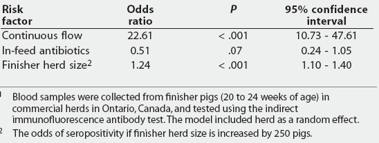
The final multivariate model (Table 1) excluded the farrow-to-finish variable because it was highly correlated with continuous flow (Pearson correlation coefficient = 0.80) and distorted the estimates of the odds ratios. Most herds with continuous-flow management in the finishing barn were farrow-to-finish herds. Herds managed by continuous flow were 23 times more likely to have seropositive pigs than herds with all-in, all-out management. When in-feed antibiotics were used in the finishing ration, herds were less likely to have seropositive pigs. The odds of a pig being seropositive increased as herd size increased by 250 finisher pigs. The H-L goodness-of-fit test suggested that the model did not fit the data very well (H-L c2 [8 df] = 78.92, P < .001).
Table 1: Logistic regression model for individual L intracellularis-seropositive status of 613 finisher pigs from 33 herds
1 Blood samples were collected from finisher pigs (20 to 24 weeks of age) in commercial herds in Ontario, Canada, and tested using the indirect immunofluorescence antibody test. The model included herd as a random effect. 2 The odds of seropositivity if finisher herd size is increased by 250 pigs. |
Discussion
The results of this study indicate that L intracellularis is widespread in the Ontario swine population, with antibodies detectable in nearly 75% of the herds tested in this study. The 90% seroprevalence in sows, regardless of parity, suggests that L intracellularis antibodies are more common in sows than in finisher pigs (seroprevalence 56%). The high seroprevalence in sows may be due to repeated exposure to L intracellularis, which would maintain antibodies at detectable levels. Loose sow housing plus floor-feeding management practices may contribute to maintaining high L intracellularis antibody levels, since contact with feces of pen-mates is very likely. Another explanation for the high seroprevalence in adult pigs might be that older animals are likely to be exposed to a greater number of microorganisms over their life spans and therefore are more likely than relatively naive young animals to possess antibodies that might cause cross-reactivity with L intracellularis. No cross reactions were reported8 in young pigs tested with the IFAT; however, in our study, adult pigs were tested, and a slightly different IFAT test, that might have caused nonspecific reactions, was used. This possibility should be investigated.
In this study, L intracellularis antibodies were detected in more than half of the finisher pigs tested. Similar results were found in a Korean study11 in which pigs from five provinces were tested with the IFAT. The proportion of positive pigs per province ranged from 43.9% to 68.6%, with an overall prevalence of 56.4%. Interestingly, L intracellularis seroprevalence was similar in wild pigs tested in the Czech Republic.12
No correlation was found between breeding-herd and finishing-herd seroprevalence. However, the Czech study12 may not have had the power to measure this association, because all breeding herds and almost all finishing herds (91%) were classified as positive. In an earlier study comparing the serological status of 123 breeding and finishing herds, the status of the breeding herd was associated with the status of the finishing herd.13 Even though the means by which L intracellularis is transmitted within a herd has not been fully studied, we believe that sows might represent a very important source of L intracellularis via fecal transmission to their offspring and from them to commingled pigs. Among 22 herds included when correlation between breeding-herd and finisher-herd prevalence was tested, finishing and breeding herds were housed at different sites in only four cases, and only one of these four finishing herds was classified as negative. The other negative finishing herd was on the same site as the breeding herd, but tylosin included in the finisher ration at 22 g per tonne might have prevented these pigs from developing a detectable immune response. From this study, it is not clear whether the number of production sites in a system affects the L intracellularis serological status of breeding and finishing herds. Breeding and finishing herds are more likely to both be positive in one-site operations because of the possibility of transmitting the organism from one group to another via carrier pigs, personnel, rodents, or fomites that move between the breeding and finishing areas. This is also likely the reason for the higher within-herd prevalence in farrow-to-finish herds compared to finishing herds.
In finishing herds stocked with feeder pigs from different sources, antibiotics are likely to be included in the feed for incoming groups as a prophylactic measure, as outbreaks of disease may occur when pigs of different immune status are commingled. It has been suggested that antibiotics may prevent infection with L intracellularis14 and antibodies are not produced, so that these pigs test seronegative. Our results corroborate this: pigs in herds using in-feed medication were less likely to be seropositive.
In this study, pigs housed in barns with continuous-flow management were more likely to be seropositive than pigs managed in all-in, all-out facilities. Continuous-flow management in finishing barns has disadvantages, compared to all-in, all-out management.15 There is a build-up of pathogens in the building when it is not completely cleaned, exposing the next group of pigs to a high concentration of pathogens that might include L intracellularis. Our results agree with those of other studies,16,17 that all-in, all-out management reduces the incidence of disease. In this study, the number of seropositive pigs was lower when all-in, all-out management was used, suggesting that the probability of exposure was lower. It is likely that on farms with all-in, all-out management, cleaning protocols are better, which tends to reduce the survivability of microorganisms. It has been suggested that strict sanitation protocols, such as cleaning, disinfection, and drying, are very important for disease control and prevention.18 If the cleaning protocol includes washing with hot water and use of disinfectants such as quaternary ammonium or iodine-based compounds,19 the bacterial challenge from L intracellularis will be decreased.
Bane et al20 found a relationship between the number of growing pigs in the herd (ie, herd size) and clinical disease, which agrees with our findings, ie, an increase in the number of finishing pigs increased the odds of a pig testing positive or being exposed to L intracellularis. In the study of Bane et al,20 the occurrence of clinical signs and histologic findings, rather than seropositivity, was the outcome measured.
We are aware that the overall fit of the model was not significant, which may be associated with the small sample size, failure to include herd in the model when the goodness-of-fit test was conducted, or both. However, we do believe that the model provides useful information.
It is important to mention that the L intracellularis avirulent live-culture vaccine was not available in Canada when these samples were collected. Therefore, in this study, antibodies induced by vaccination21 were not a confounding factor. In future studies in Ontario, antibodies produced in response to vaccination will have to be taken into account. This study suggests that L intracellularis is widespread in the swine population in Ontario, but the existing serologically negative herds will have to be cautious when introducing replacement pigs to avoid outbreaks of PPE.
Implications
- Lawsonia intracellularis infection is endemic in the province of Ontario, with almost 75% of herds having detectable levels of antibodies.
- Lawsonia intracellularis antibodies are more prevalent in sows than in finishing pigs.
- Under the conditions of this study, within-herd seroprevalence of L intracellularis is higher in farrow-to-finish farms than in multi-site operations, and in finishing barns with continuous-flow management compared to all-in, all-out management.
- The IFAT test interpreted at herd level is a good tool for determining L intracellularis herd status.
Acknowledgments
The authors thank participating producers; Marcelo Gottschalk, who performed the laboratory tests, and students and technicians who collected the samples. Funding for this project was provided by Ontario Pork, the University of Guelph-Ontario Ministry of Agriculture and Food Animal Research Program, and ELANCO Animal Health.
References
1. McOrist S, Gebhart CJ. Porcine proliferative enteropathies. In: Straw BE, D'Allaire S, Mengeling WL, Taylor DJ, eds. Diseases of Swine. 8th ed. Ames Iowa: Iowa State University Press; 1999:521-534.
*2. Gebhart C, Guedes R. Proliferative enteropathy: diagnosis and immunity. Proc AASV. 2001:353-357.
3. Holyoake PK, Cutler RS, Caple IW. A diagnostic dilemma: detecting proliferative enteritis in pigs at slaughter. Aust Vet J. 1994;71:308-309.
4. Holyoake PK, Cutler RS, Caple IW. Prevalence of proliferative enteritis on pig farms in Australia. Aust Vet J. 1994;71:418-422.
5. Wilson JB, Honour S, Pauling GE, O'Connor B, Benjamin M, Paradis MA, Dick CP. Surveillance for porcine proliferative enteropathy in Alberta by using routine diagnostic laboratory data. Can Vet J. 2002;43:604-606.
6. Wilson JB, Pauling GE, McEwen BJ, Smart N, Carman PS, Dick CP. A descriptive study of the frequency and characteristics of proliferative enteropathy in swine in Ontario by analyzing routine animal health surveillance data. Can Vet J. 1999;40:713-717.
7. Dohoo I, Martin W, Stryhn H. Sampling. In: Veterinary Epidemiologic Research. 1st ed. Charlottetown, Prince Edward Island: AVC Inc; 2003:27-52.
8. Knittel JP, Jordan DM, Schwartz KJ, Janke BH, Roof MB, McOrist S, Harris DL. Evaluation of antemortem polymerase chain reaction and serologic methods for detection of Lawsonia intracellularis-exposed pigs. Am J Vet Res. 1998;59:722-726.
9. Martin SW, Shoukri M, Thorburn M. Evaluating the health status of herds based on tests applied to individuals. Prev Vet Med. 1992;14:33-43.
10. Jordan D, McEwen SA. Herd-level test performance based on prevalence and herd true prevalence. Prev Vet Med. 1998;36:187-209.
11. Lee S, Kim T, Park S, Song CS, Chang HK, Yeh JK, Park HI, Lee JB. Prevalence of porcine proliferative enteropathy and its control with tylosin in Korea. J Vet Sci. 2001;2:209-212.
12. Tomanova K, Bartak P, Smola J. Detection of Lawsonia intracellularis in wild pigs in the Czech Republic. Vet Rec. 2002;151:765-767.
13. Bronsvoort M, Norby B, Bane D, Gardner I. Management factors associated with seropositivity to Lawsonia intracellularis in US swine herds. Swine Health Prod. 2001;9:285-290.
*14. Collins A, van Dijk N, Vu NQ, Pozo P, Love RJ. Immunity to Lawsonia intracellularis. Proc Allen D. Leman Swine Conf. 2001:115-120.
15. Harris DL, Alexander TJL. Methods of disease control. In: Straw BE, D'Allaire S, Mengeling WL, Taylor DJ, eds. Diseases of Swine. 8th ed. Ames Iowa: Iowa State University Press; 1999:1077-1110.
16. Smith SH, McOrist S, Green LE. Questionnaire survey of proliferative enteropathy on British pig farms. Vet Rec. 1998;142:690-693.
17. Stege H, Jensen TK, Moller K, Baekbo P, Jorsal SE. Risk factors for intestinal pathogens in Danish finishing pig herds. Prev Vet Med. 2001;153-164.
18. Amass SF, Clark LK. Biosecurity considerations for pork production units. Swine Health Prod. 1999;7:217-228.
19. Collins A, Love R, Pozo J, Smith S, McOrist S. Studies on the ex-vivo survival of Lawsonia intracellularis. 2000;8:211-215.
20. Bane D, Neumann E, Gebhart C, Gardner I, Norby B. Porcine proliferative enteropathy: a case-control study in the United States. Swine Health Prod. 2001;9:155-158.
21. Guedes RMC, Gebhart CJ. Onset and duration of fecal shedding, cell-mediated and humoral immune responses in pigs after challenge with a pathogenic isolate or attenuated vaccine strain of Lawsonia intracellularis. Vet Microbiol. 2003;91:135-145.
* Non-refereed references.