Case report |
Peer reviewed |
Anatomical abnormalities in a group of finishing pigs: prevalence and pig performance
Anormalidades anatómicas en un grupo de cerdos de finalización: prevalencia y desempeño del cerdo
Anomalies anatomiques dans un groupe de porcs en finition: prévalence et performances zootechniques
Barbara Straw, DVM, PhD; Ronald Bates, MS, PhD; Gerald May, MS
BS: Department of Large Animal Clinical Sciences, College of Veterinary Medicine, Michigan State University, East Lansing, Michigan. RB: Department of Animal Sciences, Michigan State University, East Lansing, Michigan. GM: Michigan State University Extension, Ithaca, Michigan. Corresponding author: Dr Barbara Straw, Large Animal Clinical Sciences, A203 Veterinary Medical Center, Michigan State University, East Lansing, MI 48824; Tel: 517-432-5199; Fax: 517-432-3450; E-mail: straw@cvm.msu.edu.
Cite as: Straw B, Bates R, May G. Anatomical abnormalities in a group of finishing pigs: prevalence and pig performance. J Swine Health Prod. 2009;17(1):28–31.
Also available as a PDF.
SummaryGrowth rate and mortality during the first 80 days in a commercial finisher were documented in pigs with scrotal or umbilical hernias or kyphosis. Umbilical hernias were classified by size. Scrotal hernias and kyphosis were not subclassified. Descriptive statistics were performed for prevalence of defects. Prevalence, gender, and mortality in affected and non-affected pigs were compared using chi-squared tests. Gain in the first 80 days was compared by ANOVA in pigs with umbilical hernias of various sizes. Prevalence and mortality rate for umbilical hernias did not differ by gender (P > .05), but kyphosis occurred more frequently in barrows (P < .05). Mortality rates were higher among affected pigs, but did not increase with umbilical-hernia score (P = .30). Pigs that died spent considerable time in the finisher, with probable compromise of their welfare during this time. Welfare and economic considerations may make euthanasia preferable to placing pigs with hernias or kyphosis in the finisher. | ResumenSe documentó el índice de crecimiento y mortalidad durante los primeros 80 días en una engorda comercial en cerdos con hernias umbilicales ó escrotales ó cifosis. Las hernias umbilicales se clasificaron por tamaño. Las hernias escrotales y cifosis no se subclasificaron. Se realizaron análisis de estadística descriptiva para la prevalencia de defectos. Se comparó la prevalencia, género, y mortalidad en cerdos afectados y no afectados utilizando la prueba de xi cuadrada. La ganancia en los primeros 80 días se comparó utilizando ANOVA en cerdos con hernias umbilicales de varios tamaños. Los índices de prevalencia y mortalidad de hernias umbilicales no difirieron por género (P > .05), pero la cifosis ocurrió más frecuentemente en machos castrados (P < .05). La mortalidad fue más altos en los cerdos afectados, pero no aumentó con la calificación de hernias umbilicales (P = .30). Los cerdos que murieron pasaron un tiempo considerable en la engorda, probablemente afectando su bienestar durante este tiempo. Las consideraciones de bienestar y económicas pueden justificar que la eutanasia sea preferible a aceptar cerdos con hernias ó cifosis en el área de finalización. | ResuméLe taux de croissance et les mortalités durant les premiers 80 jours d’élevage dans un troupeau de finition commercial ont été documentés pour des porcs ayant des hernies scrotales ou ombilicales ou de la cyphose. Les hernies ombilicales ont été classées en fonction de leur taille. Aucune sous-classification n’a été faite pour les hernies scrotales et la cyphose. Des statistiques descriptives ont été effectuées pour la prévalence des anomalies. La prévalence, le sexe, et les mortalités chez les porcs affectés et non-affectés ont été comparés à l’aide de tests de chi-carré. Le gain de poids dans les premiers 80 jours a été comparé par ANOVA chez les porcs avec hernies ombilicales de tailles différentes. La prévalence et le taux de mortalité pour les hernies ombilicales n’étaient pas différents en fonction du sexe (P > .05), mais la cyphose était plus fréquente chez les mâles castrés (P < .05). Les taux de mortalité étaient plus élevés par les porcs affectés, mais n’a pas augmenté en fonction du pointage de l’hernie ombilicale (P = .30). Les porcs qui moururent passèrent considérablement plus de temps en finition, avec fort probablement une atteinte à leur bien-être durant cette période. Pour des considérations économiques et de bien-être, l’euthanasie pourrait être préférable à l’entrée en finition pour des porcs avec hernies ou cyphose. |
Keywords: swine, scrotal hernia, umbilical hernia,
kyphosis, performance
Search the AASV web site
for pages with similar keywords.
Received: January 11, 2008
Accepted: April 23, 2008
Congenital defects occur in pigs at a prevalence estimated by different authors as 0.11% to 4.96%.1 Umbilical and inguinal hernias have been reported by one source as occurring in 0.4% to 1.5% of pigs.2 An Ontario study1 reported a prevalence of 0.39% for all types of hernias, but a higher prevalence (1.7% to 6.7%) has been reported.3 For umbilical hernias specifically, prevalence has been reported as 0.4% to 1.2%.4 For scrotal hernias, prevalence has been reported as 2% (Germany),5 5%,6 1% to 5% (Thailand),5 1.35% and 0.22% to 0.54% (Netherlands),7 and 0.6%, 1.0%, and 1.5% for the Duroc, Landrace, and Yorkshire breeds, respectively.8
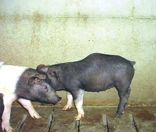
Pigs with kyphosis and lordosis are referred to in the industry as humpy-back pigs (Figure 1). In most affected animals, the condition is not apparent at birth, but becomes recognizable at 8 to 16 weeks of age, and sometimes as early as 3 weeks of age.9 The prevalence of kyphosis has been reported as 2.5% (Denmark),10 4% (England),11 and 6.3% to 11.4% (Sweden),12 with outbreaks affecting up to 30% of pigs.9
| Figure 1: Kyphosis in two pigs at placement in a
commercial finisher.
|
Identification of scrotal hernias, umbilical hernias, and kyphosis creates a dilemma for producers, as welfare and economic considerations may make euthanasia preferable to placing affected pigs in the finisher. The objective of this study was to assess growth rate and mortality of pigs with scrotal or umbilical hernias or kyphosis in a commercial finisher.
Data collection
The selected finishing site included eight 1000-head, curtain-sided, tunnel-ventilated Hog Slat barns (Hog Slat, Inc, Newton Grove, North Carolina) with totally slatted floors. Pigs were placed in the finisher at approximately 27 to 32 kg and were usually sold at 126 to 131 kg. In this observational study, approval of the study protocol by the animal care and use committee was not required. This farm employed PQA Plus guidelines13 in care of finishing pigs.
Pigs were weighed as a group at transfer to the finisher, and average weight was calculated. When the site was filled, all pigs were examined for scrotal and umbilical hernias and kyphosis, and affected pigs were individually weighed and ear-tagged, distinguishing them from the nonaffected pigs, which were not ear-tagged. Defects were assessed by a veterinarian and a group of veterinary students. Umbilical hernias were subjectively classified into three categories by approximate size: small (approximately golf-ball size), medium (approximately baseball size), and large (approximately melon size). Scrotal hernias and kyphosis were identified without further classification. Eighty days post placement, non-tagged pigs were weighed as a group and ear-tagged pigs were individually weighed. Throughout the finishing period, mortality in the tagged pigs was recorded and gross necropsies were performed on-site on all tagged and non-tagged pigs that died. Hernia contents were not cultured. Kill sheets from the packing plant were used to record mean age and individual weights of non-tagged pigs at market.
Data analysis
Descriptive statistics (prevalence of defects) and comparisons of prevalence, mortality, and gender in affected and nonaffected pigs were performed in Minitab (Minitab Inc, State College, Pennsylvania) using a chi-square test. Comparison of 80-day weight in affected and nonaffected pigs, and among pigs with small, medium, or large umbilical hernias, were performed using least squares analysis of variance (ANOVA) in SAS (SAS Institute, Cary, North Carolina). For all comparisons, the group of nonaffected pigs included only normal, healthy pigs that had not been diagnosed or treated for any diseases. For all analyses, P < .05 was considered statistically significant.
Results
No tagged pigs were euthanized during the study. No non-tagged pigs developed hernias or kyphosis after initial evaluation of the herd. Mortality rates were higher among pigs with any of the three anatomical defects than among unaffected pigs (P < .05) (Table 1). Tagged pigs that died spent up to 80 days in the finisher. Necropsy findings in pigs with hernias were characterized by peritonitis with strangulated gut. The common lesion in pigs with kyphosis was pneumonia, which was the major cause of death in this production site. Pneumonic lesions were not cultured.
Table 1: Prevalence of defects (umbilical and scrotal hernias and kyphosis) and growth rate and mortality in affected and unaffected finisher pigs*
* The 8022 pigs in the finisher were weighed as a group at placement (approximately 27 to 32 kg); 80 days later, nonaffected pigs were weighed as group, and affected pigs were individually weighed. Affected pigs were ear-tagged at placement when assessed for defects. Umbilical hernias were classified as small (approximately golf-ball size), medium (approximately baseball size), and large (approximately melon size). † Pigs not weighed at 80 days included those that died (1, 2, and 2 in the small, medium, and large umbilical hernia groups, respectively) and those that had lost their ear tags. ‡ Mean ± SD. Days until death was recorded only for pigs with hernias or kyphosis. a Differed from mortality of nonaffected pigs (chi-square analysis; P = .048). b Differed from mortality of nonaffected pigs (chi-square analysis; P < .001). c Differed from mortality of nonaffected pigs (chi-square analysis; P < .01). NA: not applicable. |
|||||||||||||||||||||||||||||||||||||||||||||||||||||
Kyphosis occurred more frequently in barrows (22) than in gilts (11) (P < .05). Among pigs with umbilical hernias, neither prevalence of the defect (P = .19) nor mortality rate (P = .41) differed between gilts (n = 36) and barrows (n = 33). No inguinal hernias were identified in gilts.
Hernias in necropsied pigs were not reducible, with some degree of fibrin adhesions evident in all cases. While the presence of an umbilical hernia was associated with slower growth rate, ADG in pigs with the largest umbilical hernias (ADG 912.0 ± 53 g) did not differ from ADG in either pigs with medium umbilical hernias (ADG 832.6 ± 30 g) or pigs with small umbilical hernias (ADG 857.9 ± 34 g; P = .43). Mortality rates were 4.0%, 3.1%, and 8.3% for pigs with umbilical hernias scored as small, medium, and large, respectively (P = .30). As the tagged pigs in this study were processed through the cull market, which provides no kill sheet, no estimate of condemnation rate was available for these pigs.
Discussion
A variety of genetic and environmental factors contribute to the formation of umbilical hernias,4 which occur when weakened supportive muscles around the umbilical stump or navel area interfere with closure of the umbilical opening, allowing intestines to protrude through the abdominal wall. The genetic control of umbilical hernias is not known. A heritable cause has been suggested,4 and progeny testing of single-sire lines showed that the odds of finding a pig with an umbilical hernia were greater for some genetic lines. However, specific genes have never been reported and umbilical hernias are not the result of simple inheritance.14 Environmental conditions that interfere with closure of the umbilical cord contribute to development of hernias, for example, abnormal stretching of the umbilical cord during farrowing, placing navel clips too close to the skin, and infection of the umbilical stump.7 Genetic variability may have an effect on the musculature of the navel, and pigs with weaker navel muscles in a poor environment may be particularly susceptible to herniation. Proper sanitation and hygiene may be more likely to reduce the incidence of umbilical hernias than eliminating certain boars or dams.
Inguinal hernias may affect both genders, although they are rare in females and usually associated with intersexuality.15 It is thought that scrotal hernias are caused by failed obliteration of the process vaginalis after descent of the testis,16 or from failed involution at the internal inguinal ring.17 In either case, the inguinal ring does not close off properly after descent of the testes, allowing the distal jejunum and ileum to drop into the scrotum. The mode of inheritance for susceptibility to inguinal and scrotal hernias is likely to be polygenic.7 In a study of breeding and performance records for an 8-year period, Vogt and Ellersieck8 identified breed differences in the prevalence of scrotal hernia in the progeny of Yorkshire, Duroc, and Landrace boars, and a greater prevalence of scrotal hernias among male full siblings of affected pigs than among male full siblings in the general population. Vogt and Ellersieck8 concluded that susceptibility to this defect is inherited via genes at multiple loci. Using a genome scan for markers associated with inguinal and scrotal hernias, Grindflek et al18 identified genomic areas associated with susceptibility to both types of hernias in pigs.
All types of hernias are classified as direct if intestines directly contact the skin, and indirect if intestinal loops outside the abdominal wall are covered by peritoneum or vaginal tunic.18 Direct contact of intestines with skin stimulates formation of adhesions that can cause partial bowel obstruction, with subsequently poor growth performance.2 The welfare of severely affected animals may be at stake if the intestine becomes completely obstructed or if the hernial sac is injured or abscessed.2 Moderate adhesions may not severely diminish performance, and the carcass values of affected and unaffected pigs should be similar. However, peritonitis interferes with evisceration at slaughter, necessitating trim loss for small hernias and, at some abattoirs, condemnation of > 50% of pigs with large hernias.2 Handling animals with hernias requires extra labor during processing, as intestinal adhesions cannot be distinguished from infectious peritonitis. Adhesions predispose to rupture of the intestines during the slaughter process, contamination of the carcass with intestinal content, and subsequent condemnation. Pigs with hernias may be marketed through specialty harvest facilities that can accommodate and slaughter them with minimal risk of carcass condemnation, as was the case in the herd observed in this study, with the caveat that special handling reduces the value of the animals.
Pigs with kyphosis may grow poorly and fail to reach slaughter weight.19 Primary vertebral lesions caused by physical or metabolic abnormalities, intrauterine infections, early onset of puberty in male pigs, stress on the lumbar spine caused by painful musculo-skeletal conditions, and genetic background have all been suggested as possible causes of kyphosis,4 but no confirmatory studies have been reported. Three variants of this defect are reported. First, there may be no gross or histological vertebral changes.10 Second, kyphosis may be the result of failure of vascularization of the ventral centers or ossification in the lumbar vertebrae, with subsequent development of ventral hemivertebrae.10 Finally, outbreaks in some herds may be associated with vasculitis affecting both the lumbar vertebrae and other tissues, as described in Canadian herds.19 An association with infectious agents such as porcine circovirus type 2 is suspected in these cases.19 Kyphosis has also been associated with lesions of osteochondrosis,10 specifically affecting intervertebral synovial joints and femorotibial joints.20 It has been suspected that osteoarthorisis and osteochondrosis may be initiated by infectious agents.21 This may include agents such as porcine reproductive and respiratory syndrome (PRRS) virus that can cross the placental barrier and infect piglets in utero.22 It has been suggested that endemic PRRS may be associated with prevalence of kyphosis in finisher pigs.9
Producers can estimate the profitability of retaining pigs with hernias or kyphosis by calculating growth performance, mortality rate, and condemnation rate for affected pigs in their herds. Welfare is an important consideration in decisions made concerning the care of these pigs. In this study, pigs with hernias not only consumed feed and occupied space in the finisher during the 3 to 4 weeks before they died, but also were likely to have experienced abdominal discomfort. Approximately 15% of pigs with hernias died during the 80-day period of observation, and previous research2 suggests that up to 50% of the survivors might have been condemned for peritonitis. Depending on availability and quality of individual-pig observation, euthanasia of affected animals when they are identified might be a better option than placing them in the finisher.
Implications
- Under the conditions of this study, mortality rates are higher in finisher pigs with umbilical or scrotal hernias or kyphosis than in unaffected animals.
- Growth rate is slower in pigs with umbilical hernias, scrotal hernias, and kyphosis than in unaffected pigs.
- Neither growth rate nor mortality rate vary with the size of an umbilical hernia.
References
1. Partlow GD, Fisher KRS, Page PD, MacMillan K, Walker AF. Prevalence and types of birth defects in Ontario swine determined by mail survey. Can J Vet Res. 1993;57:67–73.
*2. Keenliside J. Belly and scrotal ruptures (aka umbilical and inguinal hernias). 8th Ann Swine Technol Workshop. Red Deer, Alberta, Canada. 2006.
3. Thaller G, Dempfle L, Hoeschele I. Investigation of the inheritance of birth defects in swine by complex segregation analysis. J Anim Breed Genet.1996;113:77–92.
4. Searcy-Bernal R, Gardner IA, Hird DW. Effects of and factors associated with umbilical hernias in a swine herd. JAVMA. 1994;204:1660–1664.
5. Gatphayak K, Chongkasikit N, Charoensook R, Laenoi W, Vearasilp T, Sardsud V, Knorr C, ter Meulen U, Brenig B. Present situation of porcine hernia inguinalis / scrotalis in Thailand. The Global Food & Product Chain—Dynamics, Innovations, Conflicts, Strategies. Deutscher Tropentag. October 2005. Available at: http://www.tropentag.de/2005/abstracts/links/Gatphayak_F3p6xgtc.pdf. Accessed 14 July 2008.
6. Magee WT. Inheritance of scrotal hernia in swine. J Anim Sci. 1951;10:516–522.
7. Charagu PK. Congenital defects in pigs: 1. Hernias and ridglings. 2005. Available at: http://hypor.com/dbdocs//43147ed874b22.pdf. Accessed 21 July 2008.
8. Vogt DW, Ellersieck MR. Heritability of susceptibility to scrotal herniation in swine. Am J Vet Res. 1990;9:1501–1503.
*9. Sanford SE. Helping your herd get over the hump. Farm and Country Pork. June 7, 1999.
10. Nielsen LWD, Hogedal P, Arnbjerg J, Jensen HE. Juvenile kyphosis in pigs. A spontaneous model of Scheuermann’s kyphosis. Acta Pathol Microbiol Immunol (Scandinavia). 2005;113:702–707.
11. Done SH, Gresham ACJ. Lordosis and kyphosis (“humpy-back”) in pigs. Pig J. 1988;33:134–141.
*12. Bradley H. Variation in back conformation and prevalence of ulcers on the shoulders: a cohort study of related Swedish Landrace and Landrace ‘Yorkshire’ sows. University Essay from Sweden. 2005. Available at: http://www.essays.se/about/kyphosis/. Accessed 8 August 2008.
13. pork.org. Pork Quality Assurance Plus. PQA Plus Manual. Available at: www.pork.org/Producers/. Accessed 2 October 2008.
14. Rutten-Ramos SC, Deen J. Association between umbilical hernia and genetic line in a swine multiplication herd and methods to differentiate the role of sire in the incidence of umbilical hernias in offspring. J Swine Health Prod. 2006;14:317–322.
15. Tirant IN, Genghini RN, Gonzalez Quintana H, Wittouck P. Morphological and karyotypic characterization of intersex pigs with hernia inguinalis. J Agric Sci. 2002;138:333–340.
16. Clarnette TD, Lam SKL, Hudson JM. Ventriculo-peritoneal shunts in children reveal the natural history of closure of the processus vaginalis. J Pediatr Surg. 1998;33:413–416.
17. Clarnette TD, Hudson JM. Is the ascending testis actually ‘stationary’? Normal elongation of the spermatic cord is prevented by a fibrous remnant of the processus vaginalis. Pediatr Surg Int. 1997;12:155–157.
18. Grindflek E, Moe M, Taubert H, Simianer H, Lien S, Moen T. Genome-wide linkage analysis of inguinal hernia in pigs using affected sib pairs. BMC Genetics [serial online]. 2006;7:25. Available at: http://www.biomedcentral.com/1471–2156/7/25. Accessed 21 July 2008.
*19. Clark T. Hump-back pigs. Prairie Diagnostic Services and Saskatchewan Agriculture, Food and Rural Revitalization: Animal Health Expositor [serial online]. 2005;6:2–3. Available at: www.usask.ca/pds/newsletter.html. Accessed 8 August 2008.
20. Hill MA. Economic relevance, diagnosis, and countermeasures for degenerative joint disease (osteoarthrosis) and dyschondroplasia (osteochondrosis) in pigs. JAVMA. 1990;197:254–259.
21. Hill MA. Causes of degenerative joint disease (osteoarthrosis) and dyschondroplasia (osteochondrosis) in pigs. JAVMA. 1990;197:107–113.
22. Zimmerman J, Benfield DA, Murtaugh MP, Osorio F, Stevenson G, Torremorrell M. Porcine reproductive and respiratory syndrome virus (porcine Arterivirus). In: Straw B, Zimmerman J, D’Allaire S, Taylor DJ, eds. Diseases of Swine. 9th ed. Ames Iowa: Blackwell Publishing; 2006:387–418.
* Non-refereed references.