What's your interpretation?
Non Refereed
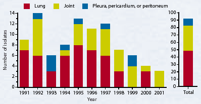
The following graph identifies the number of cases and tissues of origin from which Mycoplasma hyosynoviae was isolated from 1991 to the present at the Mycoplasma Laboratory of the Animal Health Laboratory, University of Guelph, Guelph, Ontario. Is the declining rate of isolation real? If so, is it a reflection of what is happening in the industry, or can it be explained by other factors?
Mycoplasma hyosynoviae isolated from Ontario swine
Mycoplasma hyosynoviae is one of a small group of mycoplasma organisms which, after initially colonizing the tonsil, has the ability to enter the bloodstream. It can later be isolated from various tissues, including the pericardium, lung, lymph nodes, and joints.
Is the declining rate of isolation of M hyosynoviae real?
All tissues and swabs were cultured from samples submitted for diagnostic purposes to the Mycoplasma Laboratory of the Animal Health Laboratory (AHL), University of Guelph, Guelph, Ontario (formerly the Veterinary Laboratory Services Branch, Ontario Ministry of Agriculture, Food and Rural Affairs). There were no changes in the cultural or identification techniques during this time period. Isolations were made on a modified Friis medium, with colonies identified using an immuno-fluorescent staining technique with specific M hyosynoviae antisera.
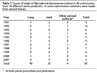
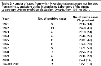
The decreased frequency of isolations of M hyosynoviae at the AHL since 1991 (Table 1 and 2) is statistically significant (Chi square for linear trend, P=.03), recognizing the submission bias to the AHL. This is, however, our best indicator so far, and it is in agreement with comments from swine practitioners.
What other factors might be involved?
The decline in the number of positive cases does not necessarily reflect the actual incidence of clinical disease in the industry, as the submission of diagnostic specimens is totally reliant on the practitioner's and producer's perception of the seriousness of the problem. Since there are typically no death losses associated with M hyosynovaie infection, is the problem severe enough to warrant submissions of diagnostic samples for which a service charge will be levied?
Management of swine operations has undergone tremendous changes in the last few years, and one can only speculate on the effect of these changes on M hyosynoviae-induced arthritis. Increased herd size, early weaning, all in-all out procedures, strict biosecurity principles, high health herds, and improved disinfection and sanitation protocols all contribute to disease incidence and expression.
In this study, as the organism was isolated more frequently from lung tissue than joints and from production units where arthritis had not been identified as a problem, it is apparent that M hyosynoviae was present in arthritis-free herds. It is also possible that arthritis was a problem in these herds, but appropriate samples were not submitted for mycoplasma isolation. Alternatively, these pigs might not have been exposed to a sufficient level of contributing factors to trigger a clinical outbreak.
At what age are pigs infected with M hyosynoviae?
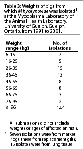 Mycoplasma hyosynoviae was
isolated from pigs of a wide range of weights, with an average
of 46.5 kg (Table 3). Several M hyosynoviae-associated
arthritis cases presented to the AHL involved fast-growing pigs.
One was a single-source, 6000-head, wean-to-finish operation,
which presented with increased numbers of pigs with bitten tails
and (or) flank sucking and (or) biting lesions. Further investigation
revealed that prior to the more obvious tail-biting, many of the
severely affected pigs had suffered from acute hind-end lameness,
with M hyosynoviae being isolated in pure culture from
affected joints. This condition had been present for several months,
with 2 to 7% of pigs per room affected. Losses were particularly
high in one of three grow-finish barns. This relatively new barn
consisted of several rooms, each holding 400 pigs. Farrowing and
nursery rooms were operated on a strictly all in-all out basis,
but grow-finish rooms were not. These pigs were noted to be extremely
restless and (or) agitated. Once roused, they would continually
pace and show aggressive behavior towards each other. In this
line of pigs, which had a very high lean growth potential, aggression
had previously been identified when a discrepancy existed between
nutrient demand and nutrient availability. This was compounded
by the co-existing presence of other factors such as subclinical
disease, ventilation inadequacy, and increased stocking density.
During periods of greatest lean deposition (grower to early finisher,
35 to 45 kg), these discrepancies were amplified, with subsequent
increased aggression, decreased rest, and, in this case, increased
occurrence of M hyosynoviae-associated arthritis.
Mycoplasma hyosynoviae was
isolated from pigs of a wide range of weights, with an average
of 46.5 kg (Table 3). Several M hyosynoviae-associated
arthritis cases presented to the AHL involved fast-growing pigs.
One was a single-source, 6000-head, wean-to-finish operation,
which presented with increased numbers of pigs with bitten tails
and (or) flank sucking and (or) biting lesions. Further investigation
revealed that prior to the more obvious tail-biting, many of the
severely affected pigs had suffered from acute hind-end lameness,
with M hyosynoviae being isolated in pure culture from
affected joints. This condition had been present for several months,
with 2 to 7% of pigs per room affected. Losses were particularly
high in one of three grow-finish barns. This relatively new barn
consisted of several rooms, each holding 400 pigs. Farrowing and
nursery rooms were operated on a strictly all in-all out basis,
but grow-finish rooms were not. These pigs were noted to be extremely
restless and (or) agitated. Once roused, they would continually
pace and show aggressive behavior towards each other. In this
line of pigs, which had a very high lean growth potential, aggression
had previously been identified when a discrepancy existed between
nutrient demand and nutrient availability. This was compounded
by the co-existing presence of other factors such as subclinical
disease, ventilation inadequacy, and increased stocking density.
During periods of greatest lean deposition (grower to early finisher,
35 to 45 kg), these discrepancies were amplified, with subsequent
increased aggression, decreased rest, and, in this case, increased
occurrence of M hyosynoviae-associated arthritis.
The producer's solution was novel, considering the physical and operational constraints. Pulse medication with antibiotics met with only limited success. To counteract the aggressive nature of these pigs, on a daily rotating basis, he allowed one pen of pigs access to the central alleyway. Growth parameters were no doubt negatively affected, but tail biting and arthritis virtually stopped.
To the already long list of factors that contribute to clinical expression of arthritis, can we now add a genetic component that would encompass factors other than conformation and poor leg action?
-- Gaylan Josephson, DVM, DipPath
-- Beverly McEwen, DVM, MSc, PhD, Diplomate ASVP
-- Tony van Dreumel, DVM,MSc, Diplomate ASVP
-- Pat McRaild
Animal Health Laboratory,
University of Guelph,
Guelph, Ontario.