| Original research | Peer reviewed |
SummaryObjectives: To evaluate the effectiveness of disinfection protocols on porcine circovirus type 2 (PCV2) in 1:61 scale model livestock trailers and determine the infectivity of PCV2 DNA detected using a bioassay. Materials and methods: Each model trailer was contaminated with intestinal content from a pig experimentally infected with PCV2, washed, and disinfected with one of four protocols. Between protocol steps, swabs were collected from six different areas of each trailer and tested for PCV2 DNA. Disinfection protocols were quaternary ammonium compound (DISF1), oxidizing agent containing potassium peroxomonosulfate (DISF2), combined glutaraldehyde and quaternary ammonium product (DISF3), and DISF2 followed by sodium hypochlorite compound (DISF4). Four PCV2-contaminated, nonwashed, nondisinfected trailers served as positive controls and an uncontaminated trailer as negative control. Two 6- to 10-week-old PCV2-naive pigs were placed in each cleaned and disinfected trailer for 2 hours. Blood samples collected weekly for 7 weeks were tested for anti-PCV2 IgG antibodies and PCV2 DNA. Results: In all four disinfection protocols, the amount of PCV2 DNA was significantly smaller (P < .05) after the initial wash; however, only in DISF4 (P < .01) was the amount of PCV2 DNA reduced after the disinfection step. After disinfection, 1.5 to 5.3 log10 PCV2 genomic copies per mL were identified in the trailers. While animals exposed to positive control trailers became viremic and seroconverted, no seroconversion or viremia was detected in animals exposed to disinfected trailers. Implications: Although detectable amounts of PCV2 may remain in transport vehicles after proper disinfection, risk of transmission appears minimal. | ResumenObjetivos: Evaluar la eficacia de los protocolos de desinfección contra circovirus porcino tipo 2 (PCV2 por sus siglas en inglés) en camiones en un modelo a escala 1:61 y determinar la infectividad del DNA de PCV2 detectado utilizando bioensayo. Materiales y métodos: Se contaminó cada modelo de camión con contenidos intestinales de un cerdo infectado experimentalmente con PCV2, se lavó, y se desinfectó con uno de cuatro protocolos. Entre las etapas de cada protocolo, con hisopos, se recolectaron muestras de seis diferentes áreas de cada camión y se analizaron en busca de DNA de PCV2. Los protocolos de desinfección utilizados fueron cuaternarios de amonio compuesto (DISF1 por sus siglas en inglés), un agente oxidante que contenía peroxomonosulfato de potasio (DISF2 por sus siglas en inglés), un producto combinado con glutaraldehido y cuaternario de amonio (DISF3 por sus siglas en inglés), y el DISF2 seguido de compuesto de hipoclorito de sodio (DISF4 por sus siglas en inglés). Cuatro camiones contaminados con PCV2, no lavados, no desinfectados, sirvieron como controles positivos y un tráiler no contaminado como control negativo. Dos cerdos de 6 a 10 semanas de edad libres de PCV2 se colocaron durante dos horas en cada camión lavado y desinfectado. Se analizaron las muestras de sangre colectadas semanalmente durante 7 semanas en busca de anticuerpos IgG anti PCV2 y DNA de PCV2. Resultados: En los cuatro protocolos de desinfección, la cantidad de DNA de PCV2 fue significativamente menor (P < .05) después del lavado inicial; sin embargo, sólo en el grupo DISF4 (P < .01) la cantidad de DNA de PCV2 se redujo después de la etapa de desinfección. Después de la desinfección, se detectaron entre 1.5 a 5.3 log10 copias genómicas de PCV2 por mL en los camiones. Mientras que los animales expuestos a los camiones del grupo control positivo se volvieron virémicos y seroconvertieron, no se detectó viremia ó seroconversión en animales expuestos a los camiones desinfectados. Implicaciones: A pesar de que puedan quedar cantidades detectables de PCV2 en los camiones de transporte después de una desinfección adecuada, el riesgo de transmisión es mínimo. | ResuméObjectifs: Évaluer l’efficacité de protocoles de désinfection contre le circovirus porcin de type 2 (PCV2) dans des remorques à bestiaux à l’échelle 1:61 et déterminer l’infectivité de l’ADN de PCV2 détecté à l’aide d’un bio-essai. Matériels et méthodes: Chaque modèle de remorque a été contaminé avec du contenu intestinal provenant d’un porc infecté expérimentalement avec PCV2, lavé, et désinfecté avec l’un des quatre protocoles. Entre les étapes des protocoles, des écouvillons ont été prélevés de six endroits différents dans chaque remorque et testés pour l’ADN de PCV2. Les différents protocoles de désinfection étaient composé d’ammonium quaternaire (DISF1); agent oxydant contenant du peroxomonosulfate de potassium (DISF2); un produit combinant du glutaraldéhyde et un ammonium quaternaire (DISF3); et DISF2 suivi d’un composé d’hypochlorite de sodium (DISF4). Quatre remorques contaminées avec PCV2 mais non-lavées et non-désinfectées ont servi de témoins positifs et une remorque non-contaminée de témoin négatif. Deux porcs naïfs pour le PCV2, âgés de 6 à 10 semaines, ont été placés dans chaque remorque lavée et désinfectée pour une période de 2 heures. Des échantillons sanguins prélevés de manière hebdomadaire pendant 7 semaines ont été testés pour détecter des anticorps de type IgG anti-PCV2 ainsi que de l’ADN de PCV2. Résultats: Pour les quatre protocoles de désinfection, la quantité d’ADN de PCV2 était significativement inférieure (P < .05) après le lavage initial; toutefois, seulement lors de l’utilisation du DISF4 (P < .01) a-t-on pu noter une réduction de l’ADN de PCV2 après l’étape de la désinfection. Après la désinfection on trouva de 1.5 à 5.3 log10 copies du génome de PCV2 par mL dans les remorques. Alors que les animaux exposés aux remorques témoins positifs devinrent virémiques et ont séro-convertis, aucune séro-conversion ou virémie ne fut détectée chez les animaux exposés aux remorques désinfectées. Implications: Bien que des quantités détectables de PCV2 peuvent demeurer dans les véhicules de transport suite à une désinfection appropriée, le risque de transmission apparaît minime. |
Keywords: swine, porcine circovirus type 2, transmission, transportation, PCV2
Search the AASV web site
for pages with similar keywords.
Received: May 30, 2010
Accepted: October 1, 2010
Porcine circovirus (PCV) type 2 (PCV2) is a small, non-enveloped, circular, single-stranded DNA virus that was first isolated from pigs exhibiting multisystemic wasting disease in the late 1990s.1 Since the initial description, PCV-associated disease (PCVAD) has been reported in most pig-producing countries worldwide, and the virus is now considered ubiquitous.2 Porcine circovirus associated disease is now manifested in a variety of ways, including multisystemic disease with wasting, reproductive failure, enteritis, respiratory disease, porcine dermatitis and nephropathy syndrome, or a combination of these.3 In cases of PCVAD, a morbidity rate of 4% to 30%, with a mortality rate of 70% to 80% in affected animals, has been reported.4 Commercial PCV2 vaccines are now available and have generally been very effective in reducing morbidity and mortality in the field5 and under experimental conditions.6
Porcine circovirus type 2 is shed in respiratory secretions, oral secretions, urine, and feces.7 It is a hardy virus that is quite stable in the environment. In an in vitro study,8 PCV2 virus stock was subjected to various heat treatments ranging from 56°C to 85°C for 15 minutes. Following incubation, the virus stock was added to PK-15 cells and evidence of PCV2 infection was documented. Under these conditions, PCV2 antigen staining was not detected at treatment temperatures between 75°C and 85°C, although PCV2 polymerase chain reaction (PCR) products were identified in samples treated at 75°C. In other studies, dry-heat treatment of freeze-dried PCV2 at 120°C for 30 minutes resulted in only a 1-log reduction of the virus,9 and PCV2 retained an infectivity titer of 1 × 102.7 following exposure to a pH of 2 for 30 minutes, and a titer of 1 × 102.5 following incubation at 70°C for 1 hour.10
Previous work also suggests that under in vitro conditions, mean PCV2 titers were reduced but not completely eliminated after exposure to various disinfectants, with oxidizing products and aldehyde-quaternary ammonium combination products showing the highest efficacy.11-13 In a separate study,10 exposure of PCV2 cell suspensions to an oxidizing product or to a sodium hydroxide compound led to a complete reduction in PCV2 infectivity after a 10-minute incubation period. However, neither a cytotoxicity control nor a virus control (virus plus medium alone) were used in this study. While the above information exists on the effectiveness of disinfectants under in vitro conditions, to the authors’ knowledge, no information exists on the effectiveness of disinfectants under field conditions.
Knowledge of the modes of transmission of PCV2 is vital in understanding the ecology, epidemiology, and control of PCVAD.14 Specific information on the horizontal transmission of PCV2 via fomites such as commercial livestock transport trailers is lacking. The increased use of multi-site production facilities has led to an increased frequency of animal movement, and with this, there is an increased risk of pig exposure to pathogens while in transport. In addition, the transmission of other pathogens, including Salmonella, Escherichia coli, and porcine reproductive and respiratory syndrome virus (PRRSV), through contaminated transport trailers has previously been investigated. Washing and sanitizing trailers significantly decreased the numbers of Salmonella and E coli bacteria present in trailers.15 Previous work on PRRSV has also shown that washing combined with the use of an efficacious disinfectant for 90 to 120 minutes, applied as a foam, likely reduces the number of infectious virus particles in model trailers.16 Similarly, fogging model trailers with a glutaraldehyde-quaternary ammonium chloride product (30 minutes exposure time) or an 8-hour period of drying at 20°C resulted in little (two of 19 samples) or no (none of 20) detection of PRRSV by PCR.17 Model trailers have also been used to evaluate thermo-assisted drying and decontamination for elimination of PRRSV.18 The use of a model-trailer protocol to evaluate transmission of PRRSV in previous studies provides validation for the use of this model in the study of PCV2 transmission. Accordingly, the protocol used in this study is similar to that described in previous work on PRRSV.16-18
To the authors’ knowledge, no information exists to date on the ability of commercial transport trailers to aid in transmission of PCV2 or on the ability of various disinfectant protocols to decrease or eliminate the risk of transmission of PCV2 via this route. The first objective of this study was to evaluate the ability of four disinfection protocols with known in vitro effectiveness against PCV2 to prevent transmission of PCV2 under simulated field conditions. To accomplish this, the amount of PCV2 DNA present in PCV2-contaminated 1:61 scale model trailers, washed and then disinfected with four different protocols, was compared to the amount of PCV2 DNA present in a PCV2-contaminated, nonwashed, nondisinfected model trailer. The second objective of this study was to determine the infectivity of the PCV2 DNA detected by PCR. For this, naive pigs were exposed to model trailers either disinfected with four different disinfectant protocols or subjected to two control treatments.
Materials and methods
The experimental protocol in this study was approved by the Iowa State University Institutional Animal Care and Use Committee.
Experimental design
The first objective of the study was to determine if the disinfection step reduced the amount of PCV2 present on the model trailer surface. To accomplish this, three trailer models (experimental units: Figure 1A) were either contaminated with fecal material collected from a pig experimentally infected with PCV2 and showing clinical signs consistent with PCVAD (n = 11) or remained uncontaminated (n = 1). Six swabs (subsamples) were collected from each model trailer immediately after contamination (within 10 minutes), designated as “post-contamination” samples, and tested for the presence of PCV2 DNA by real-time PCR.19 Subsamples were collected from six different model-trailer surface locations, including the aluminum smooth-sheet side wall, a hinged aluminum simulated roll-up rear door, the aluminum alloy diamond-plate flooring, a latch, an overlapped seam at the side post, and the center dividing gate (Figure 1B-1G). After contamination with PCV2, positive control model trailers (n = 4) were not washed. All other contaminated model trailers (n = 7) were washed. Six swabs of each of the washed model trailers were collected, designated as “post-wash,” and tested by real-time PCV2 PCR.19 After the wash step, each model trailer was disinfected with one of four disinfection protocols (DISF1 through DSF4). Six swabs of each of the disinfected model trailers were collected, designated as “post-disinfection,” and tested by PCV2 real-time PCR.19 A summary of the number of replicates and swabs collected at each sampling point is outlined in Table 1. High numbers of replicates of the positive control treatment were used to gain a statistically more precise estimate of a subset of the collected data (post contamination versus post wash, and post contamination versus post disinfection).
Figure 1: A: 1:61 scale model of a full-sized standard livestock trailer used in the transportation of commercial swine; B: Models were composed of aluminum smooth-sheet side walls welded and riveted to tubular extruded aluminum cross-members; C: Hinged aluminum simulated roll-up rear door; D: Aluminum alloy diamond-plate flooring; E: Latch; F: Overlapped seam at the side post; and G: Center divide gate. Dimensions were 0.62 m width × 0.82 m height × 1.11 m length. 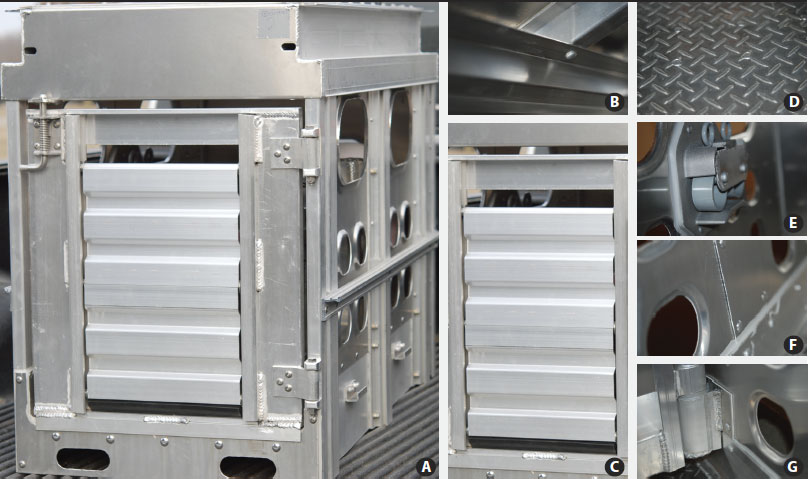 |
Table 1: Evaluation of four disinfection methods* for their ability to reduce the amount of porcine circovirus type 2 (PCV2) in seven 1:61 scale model livestock trailers contaminated with intestinal content collected from a pig experimentally infected with PCV2 then washed and disinfected†
* DISF1, a quaternary ammonium compound (PI Quat 20; Preserve International, Reno, Nevada); DISF2, an oxidizing agent containing a combination of potassium peroxymonosulfate and sodium chloride (VirkonS; DuPont Chemical Solutions Enterprise, Wilmington, Delaware); DISF3, a combined glutaraldehyde and quaternary ammonium product (Synergize; Preserve International); and DISF4, an application of two disinfectants in tandem: first, an oxidizing agent (VirkonS; DuPont Chemical Solutions Enterprise); second, a chloride compound (Clorox Bleach; The Clorox Company, Oakland, California). Each disinfectant was used as directed by the manufacturer. † After contamination, washing, and disinfection steps, swabs were collected from six designated sites in each trailer and tested by polymerase chain reaction for PCV2. Mean and SE of log10 genomic copies/mL of PCV2 DNA are reported from each of the three sampling times (post contamination, post wash, post disinfection). Data from swabs collected after the post-contamination step or the post-wash step were considered to be part of a single group in each case, regardless of the disinfectant used in the succeeding step. Post-disinfection samples were considered independently for each disinfection protocol. ‡ There was one replicate for the DISF4 treatment because, on the basis of previous work,11,12 it was expected to be most different from the positive control. abc Different superscripts within a column indicate significant differences among steps (P < .05; Tukey-Kramer-adjusted t value for differences of least squares means). |
||||||||||||||||||||||||||||||||||||||||
The second objective of the study was to determine whether the PCV2 DNA present in disinfected model trailers (DISF1 through DISF4) was infectious to naive animals. To answer this question, a total of 24 PCV2-naive pigs were placed in groups of two in model trailers that were randomly assigned to a treatment (positive control, n = 8 pigs; negative control, n = 2 pigs; DISF1, n = 4 pigs; DISF2, n = 4 pigs; DISF3, n = 4 pigs; DISF4, n = 2 pigs). The pigs were exposed to the model trailers in blocks of six (two pigs in each of three trailer models; three treatment groups tested per block) for a total of 2 hours. The time of exposure was as previously described in PRRSV transmission studies.16-18 After this, the pigs were moved to the research facility and were housed by group in rooms for 49 days following model trailer exposure. All pigs were monitored on a weekly basis for development of anti-PCV2 antibodies by ELISA20 and for PCV2 viremia by real-time PCR.19
For this portion of the study, an incomplete block design was used, with three experimental units per block (model trailers containing two pigs), four blocks, and six treatments (Table 2). Within each block, the model trailer was considered the independent experimental unit and treatments were randomly assigned to trailers. Each block included a positive control treatment and two of the remaining five possible treatments (DISF1 through DISF4 or negative control). There were two replicates each for the DISF1, DISF2, and DISF3 treatments. There was one replicate each for the negative control and DISF4 treatments, because those treatments were expected to be the most different from the positive control as determined by previous in vitro work.11,12 Blocks were included in the study to account for variation in environmental conditions.
Table 2: Incomplete block design using three model trailers and four replicates to determine whether PCV2-naive animals would become PCV2-seropositive or viremic following a 2-hour exposure period to PCV2-contaminated, nonwashed, nondisinfected trailers (positive control treatment; POS); PCV2-contaminated, washed, disinfected trailers (DISF1 through DISF4 treatments*), or uncontaminated trailers (negative control treatment; NEG)
* Disinfectant treatments described in Table 1. |
PCV2-contaminated fecal material
A colostrum-fed, crossbred, specific-pathogen-free, PCV2-naive pig was inoculated with 5 mL (3 mL intranasally and 2 mL intramuscularly) of 104.0 50% tissue culture infectious doses (TCID50) per mL of PCV2b isolate NC-16845 (accession no. EU340258) at 3 weeks of age. At 35 days post inoculation, the pig was euthanized with an overdose of pentobarbital (Fatal Plus; Vortech Pharmaceutical, Dearborn, Michigan) due to the development of clinical signs consistent with PCVAD, including severe dyspnea, diarrhea, and loss of condition. After euthanasia, the complete contents of the small intestine, cecum, and colon were collected. Twenty-mL aliquots of intestinal content were transferred into 50-mL centrifuge tubes and stored at -80°C until use. A portion of the fecal material was tested for the presence of PCV2 DNA by real-time PCR;19 10.8 log10 genomic copies per mL of PCV2 DNA were recovered. Systemic infection of the animal with PCV2 was further confirmed by the presence of intense PCV2-antigen staining by immunohistochemistry (IHC)21 in the lungs, intestine, and lymphoid tissues. Transmissible gastroenteritis virus antigen, rotavirus type 2 antigen, and Lawsonia intracellularis antigen were not detected by IHC. PRRS virus RNA, swine influenza virus (SIV) RNA, and Mycoplasma hyopneumoniae DNA were not detected by PCR. All tests were performed according to the standard operating procedures for the specific tests at the Iowa State University Veterinary Diagnostic Laboratory.
Trailers
For each block, three model trailers were used to evaluate three of the six treatments. Trailer models were designed and constructed by a company specializing in standard and custom aluminum livestock transportation equipment (EBY Inc, Story City, Iowa). The trailer models were specifically designed to accurately represent a standard livestock trailer used in the transportation of commercial swine and provided adequate space to allow animals to turn completely around. The models were composed of 29-mm thick 6061 T-6 aluminum alloy diamond-plate flooring welded and riveted to tubular extruded aluminum cross-members; 19-mm thick aluminum smooth-sheet side walls with both large (25.4 cm × 14.9 cm) and small (8.9 cm × 7.0 cm) ventilation holes with an overlapped seam at the side post; extruded aluminum side posts with integral closure track; a hinged aluminum rear door; a center divide gate; and a simulated roll-up door (Figure 1). Final dimensions were 0.62 m (width) × 0.82 m (height) × 1.11 m (length). The trailer models were 1:61 scale models of a full-sized trailer and had a floor space of 0.69 m2, which corresponded to 41.85 m2 (2.59 m wide × 16.16 m long) for the full-sized trailer, and a constant animal density of 0.34 m2 (trailer floor space in m2 divided by number of animals per trailer).
Trailer sampling protocol
At each of the four sampling points (precontamination, post contamination, post wash, post disinfection), six swabs (polyester-tipped swab; Fisher Scientific Inc, Pittsburgh, Pennsylvania) were collected from each model trailer. Designated areas included the aluminum smooth-sheet side wall, the hinged aluminum simulated roll-up rear door, the aluminum alloy diamond-plate flooring, a latch, an overlapped seam at the side post, and the center dividing gate (Figure 1B-1G). Each swab was placed into 1 mL of sterile saline (0.9% sodium chloride solution; Fisher Scientific Inc), used to swab a 5.5-cm diameter area (using a sterile template), and then stored at -80°C in a 5-mL plastic tube (BD falcon tube; Fisher Scientific Inc) prior to testing by PCR. To prevent carryover contamination between model trailers, a new template was used for each trailer, and new gloves were used by the investigator. To prevent carryover between sampling steps, templates were submerged in an oxidizing disinfectant (Virkon S; DuPont Chemical Solutions Enterprise, Wilmington, Delaware) for 10 minutes between sampling points.
Trailer contamination protocol
To contaminate each of the trailers (except the trailer used for the negative control treatment), a 20-mL aliquot of the PCV2-contaminated fecal material was thawed at 4°C overnight and spread on the walls and floor of the trailer by hand by the same investigator for each trailer within a block. After contamination, 200 g of bedding (wood chips; Theisen’s, Ames, Iowa) were placed on the floor of the trailer. In order to maintain similar experimental conditions between groups, bedding was also placed in the trailer assigned to the negative control treatment.
Trailer washing protocol
To prevent cross-over contamination from the previous step, investigators changed gloves prior to the wash step. The trailers were washed using the following protocol. The trailer was first rinsed using a low-pressure nozzle (50 psi) to flush out the majority of woodchips and gross debris, then washed with 48.9°C water using a high-pressure nozzle (2400 psi) for approximately 30 seconds, and last rinsed using a low-pressure nozzle to remove any remaining gross debris.
Trailer disinfection protocol
To prevent crossover contamination from the previous step, a different investigator or an investigator who had showered and changed clothes and gloves applied disinfectant to the trailer. Gloves were changed between trailers, and ample room was available to prevent crossover of disinfectants between trailers within a block. Disinfectants were applied using a liquid concentrate sprayer (H. D. Hudson Manufacturing Company, Chicago, Illinois) attached to a garden hose. The following disinfectants, which correspond to the experimental design in Table 1, were used: a quaternary ammonium compound (PI Quat 20; Preserve International, Reno, Nevada) (DISF1); an oxidizing agent containing a potassium peroxomonosulfate (Virkon S) (DISF2); a combined glutaraldehyde and quaternary ammonium product (Synergize, Preserve International, Reno, Nevada) (DISF3); and an application of two disinfectants in tandem, the first being the previously described oxidizing agent (Virkon S) and the second a chloride compound (Clorox Bleach; The Clorox Company, Oakland, California) (DISF4). Treatments were applied at the following rates, based on the respective manufacturers’ recommendations: DISF1 was applied at a rate of 2 oz per 5 gal of water (59 mL per 18.9 L), DISF2 was reconstituted to a concentration of 100 g per L of water and applied at a rate of 1 oz per gal (30 mL per 3.8 L), and DISF3 was applied at a rate of 0.5 oz per gal of water (15 mL per 3.8 L). For DISF4, the oxidizing agent was applied at the previously described rate and the chloride compound was applied at a rate of 6 oz per gal of water (177.4 mL per 3.8 L). Disinfectants were applied, and after 10 minutes of contact time, the trailers were rinsed with water. The temperature during the disinfectant process was 20°C for all blocks.
Decontamination protocol between blocks
To ensure that residual contamination of each trailer between the four blocks did not occur, trailers were washed with 48.9°C water using a high-pressure nozzle (2400 psi), scrubbed with a detergent product (Ultra Palmolive OXY Plus with Bleach Alternative; Colgate-Palmolive Company, New York, New York), thoroughly rinsed, disinfected with an oxidizing agent (VirkonS), and allowed to dry completely. This process was repeated until PCV2 DNA was not detected by real-time PCR from swabs collected from six different trailer surface locations (Figure 1B-1G).
Animals
Blocks 1 and 2 were performed with animals from source A in September 2008, and Blocks 3 and 4 were performed using pigs from source B in February 2009. Obtaining pigs from two sources was not part of the study design, but it was necessary due to the inability to obtain PCV2-naive animals from source A in February of 2009. The use of blocks accounted for the variability introduced when two pig sources were used. In both sources, pigs were colostrum-fed, crossbred, specific-pathogen-free conventional pigs purchased from herds that were routinely tested for major swine pathogens and known to be free of PCV2, PRRSV, porcine parvovirus, and SIV. Source A pigs were weaned at 3 weeks of age and raised to approximately 18 to 27 kg (8 to 10 weeks old), at which time they were transported to the Livestock Infectious Disease Isolation Facility (LIDIF) at Iowa State University, Ames, Iowa. Source B pigs were weaned at 2 weeks of age and raised to approximately 10 to 22 kg (6 to 9 weeks old), at which time they were transported to the LIDIF. Pigs were housed in groups of two in separate rooms with dedicated ventilation systems. Each room had 18 m2 of solid concrete floor space and one nipple drinker. Once a day, all groups were fed a balanced, pelleted, complete-feed ration free of animal proteins (excluding whey) and antibiotics (Nature’s Made; Heartland Co-op, Iowa). Upon arrival at the facility, serum from each pig was tested by real-time PCV2 PCR19 and PCV2 ELISA20 to confirm that all were PCV2-free prior to initiation of the trial.
Exposure of the pigs to the trailer models
After disinfection, the three model trailers were placed into a clean equine trailer (no prior exposure to swine) and separated by clear polyethylene sheeting. Two pigs were placed in each model trailer for a total exposure time of 2 hours. The 2-hour exposure period was similar to that used in previously published studies utilizing model trailers.16-18 During the exposure time, the horse trailer containing the model trailers was driven on a specific route, which included approximately 32.2 km of interstate highway, 56.3 km of state highway, and 32.2 km unpaved (gravel) road through Hardin, Story, and Hamilton counties in the state of Iowa. After the 2-hour exposure period, pigs were placed in isolation rooms by treatment group. At the time when Block 1 was being conducted, the temperature was 22.8°C and it rained throughout the time of transport. For Block 2, the temperature was 29.4°C with no precipitation. For Block 3, the temperature was 1.1°C with no precipitation. For Block 4, the temperature was -12.2°C with no precipitation.
Anti-PCV2 IgG antibodies and PCV2 DNA quantification and sequencing
Blood samples were collected on the day of exposure and weekly thereafter until 49 days post trailer exposure. The blood was collected in 8.5-mL serum separator tubes (Fisher Scientific Inc, Fair Lawn, New Jersey), immediately centrifuged at 2000g for 10 minutes at 4°C, and stored at -80°C until use. Serum samples were tested by an open reading frame 2 (ORF2)-based PCV2 IgG ELISA and were considered positive if the calculated sample-to-positive (S:P) ratio was ≥ 0.2, as previously described.20 Deoxyribonucleic acid extraction on serum samples was performed using the QIAamp DNA Mini Kit (Qiagen Inc, Valencia, California). Previously described primers for ORF1 of PCV219 were used for quantification of the PCV2 genomic DNA by real-time PCR. The PCR reaction consisted of 25 µL PCR mixtures that contained 12.5 µL of commercially available master mix (TaqMan Universal PCR Master Mix; Applied Biosystems Inc, Foster City, California), 2.5 µL DNA extract, and 1 µL forward and reverse primers and 0.5 µL detection probe with concentrations of 10 µM each. On each plate, five progressive 1:10 dilutions of a known copy number of PCV2 genomic DNA excised from a purified PCV2 DNA clone was included to generate a standard curve. Each plate was run in the sequence detection system (7500 Sequence Detection System; Applied Biosystems Inc) under the following conditions: 2 minutes at 50°C, 10 minutes at 95°C, followed by 40 cycles of 15 seconds at 95°C and 1 minute at 60°C. Samples which did not generate a signal after 40 cycles were considered negative. Polymerase chain reaction products amplified from virus recovered from a serum sample from one animal in each of the positive control groups was sequenced and compared with the PCV2 in the fecal material used to contaminate the model trailers. A nested PCR was used to amplify the entire ORF2 gene for sequencing as previously described.22 Polymerase chain reaction products were purified using the QIAquick PCR purification kit (Qiagen Inc) per the manufacturers’ directions and sequenced at Iowa State University DNA facility. Sequences were analyzed with Sequence Scanner 1.0 (Applied Biosystems Inc) and compared with the sequence of the PCV2 present in the fecal material using the basic local alignment search tool (BLAST).23
Necropsy
All pigs were necropsied 49 days post model-trailer exposure. Severity of macroscopic lung lesions, ranging from 0% to 100% of the lung affected, were estimated subjectively, and the size of lymph nodes was scored from 0 (normal) to 3 (four times the normal size) in a blinded fashion as described previously.24 Sections of lymph nodes (superficial inguinal, mediastinal, tracheobronchial, mesenteric), tonsil, thymus, ileum, kidney, colon, spleen, and liver were collected at necropsy, fixed in 10% neutral-buffered formalin, and routinely processed for histological examination.
Histopathology
Microscopic lesions were evaluated by a pathologist blinded to treatment groups. Lung sections were scored for the presence and severity of interstitial pneumonia ranging from 0 (normal) to 4 (severe interstitial pneumonia) as described previously.25 Sections of heart, liver, kidney, ileum, and colon were evaluated for lymphohistiocytic inflammation and scored from 0 (none) to 3 (severe). Lymphoid tissues, including lymph nodes, tonsil, and spleen, were evaluated for lymphoid depletion ranging from 0 (normal) to 3 (severe) and histiocytic inflammation and replacement of follicles ranging from 0 (normal) to 3 (severe).26
Immunohistochemistry
Immunohistochemistry for detection of PCV2-specific antigen was performed on formalin-fixed and paraffin-embedded sections of lymph nodes (superficial inguinal, mediastinal, tracheobronchial, and mesenteric), tonsil, spleen, Peyer’s patches, and thymus using a rabbit polyclonal antiserum as described previously.21 Tissues were scored for PCV2 antigen by a pathologist blinded to treatment groups. Scores ranged from 0 (no signal) to 3 (more than 50% of the lymphoid follicles contain cells with PCV2 antigen staining).26
Statistical analysis
The statistical analysis for the first objective, to determine if disinfection reduced the amount of PCV2 present on the model trailer surface, focused on the differences in numbers of log10 genomic copies of PCV2 between steps of each treatment protocol (post contamination, post washing, and post disinfection) and among four disinfectants. The experimental unit was the model trailer within a block. The assumption of independence of experimental units was reasonable because each model trailer was carefully cleaned to eliminate carryover between blocks. The amount of PCV2 DNA was measured on six swabs (subsamples) at each step within an experimental unit; these were averaged before statistical analysis.
Model trailers assigned to DISF1 through DISF4 treatments were sampled at four steps of the decontamination process (precontamination, post contamination, post washing, and post disinfection). Model trailers assigned to the positive control treatment were sampled only twice (precontamination and post contamination). Model trailers assigned to the negative control treatment were sampled only once. Data from the precontamination step and the negative control treatment were excluded from the analysis because no PCV2 DNA copies were detected. The positive control and DISF1 through DISF4 treatments were used to define three unique sampling steps: post contamination (positive control treatment and DISF1 through DISF4), post washing (DISF1 through DISF4 combined), and individual post-disinfection treatments (DISF1, DISF2, DISF3, or DISF4). This is appropriate because after contamination (prior to washing), all trailers were treated alike, and after washing (prior to disinfection), all washed trailers were treated alike.
The statistical analysis accounted for correlation between the three repeated measurements on the disinfected trailers (post contamination, post washing, and post disinfection) by including a random effect for the experimental unit. Differences in the average log10 genomic PCV2 copies per mL among the treatment groups were tested using PROC MIXED in SAS version 9.2 (SAS Institute, Inc, Cary, North Carolina), based on a linear regression model. The variation in environmental temperatures was accounted for by including block as a fixed effect in the model. Variance components were estimated using the restricted maximum likelihood (REML), and the Satterthwaite approximation was used to compute degrees of freedom. The type 3 F test of treatments was considered significant if the P value was < .05. Tukey-Kramer-adjusted t values were determined if the overall F test was significant.
There was no statistical evaluation for the second objective of the experiment, as neither seroconversion nor viremia was detected in any of the animals exposed to disinfected model trailers (DISF1 through DISF4).
Results
Amount of PCV2 DNA on the model trailer surfaces was reduced after the wash step. The mean amounts of PCV2 DNA (log10 genomic copies per mL) obtained for each of three sampling points (post contamination, post wash, and four levels of post disinfection representing the four disinfectant protocols) are presented in Table 1. Overall, the log10 PCV2 copies per mL differed by treatment (P < .001). Differences of least squares means using the Tukey-Kramer-adjusted t test indicated a significant reduction in mean log10 genomic copies per mL of PCV2 DNA between the post-contamination step and the post-wash step. Only in the DISF4 treatment group was the mean amount of PCV2 DNA significantly less (P < .01) after the disinfection step than it had been after the post-wash step.
Evidence of PCV2 infection was not present in naive animals exposed to contaminated, disinfected model trailers. Clinical disease was not observed in any of the pigs for the duration of the study. Seroconversion to PCV2 was not detected in any of the animals exposed to the negative control, DISF1, DISF2, DISF3, or DISF4 treatments during any of the replicates. Both positive control pigs seroconverted by 28 days post model-trailer exposure in blocks 1, 2, and 4. In block 3, one positive control pig seroconverted by 14 days post model-trailer exposure and the other by 42 days post exposure. Viremia was not detected in any of the animals exposed to trailers treated with DISF1, DISF2, DISF3, or DISF4 protocols or the negative control treatment during any of the replicates. Both pigs in the positive control group became PCV2 viremic by 21 days post exposure in blocks 1, 3, and 4. Both positive control pigs in block 2 became PCV2 viremic by 14 days post exposure. Characteristic microscopic lesions of PCV2 infection or PCV2 antigen staining were not detected in any of the animals from the negative control, DISF1, DISF2, DISF3, or DISF4 groups. In positive control pigs (n = 8), there was mild to moderate depletion of follicles in the tonsil (one of eight) and in lymph nodes (five of eight). Histiocytic replacement of lymphoid germinal centers was observed in two of eight animals. Low to moderate amounts of PCV2 antigen were detected in the tonsils of four of the eight pigs and in lymph nodes of six of the eight pigs. Mild lymphoplasmacytic myocarditis, hepatitis, and interstitial nephritis were noted in three of eight, two of eight, and three of eight animals, respectively.
Discussion
The first objective of this study was to evaluate the ability of four disinfection protocols with known in vitro effectiveness against PCV2 to prevent transmission of PCV2 under simulated field conditions, and the second objective was to determine the infectivity of the PCV2 DNA detected by PCR. Previous work has shown that oxidizing agents, aldehyde compounds, and quaternary ammonium compounds have efficacy against PCV2 under in vitro conditions.10-12 While in vitro efficacy data is useful, field efficacy of a disinfectant depends on the type of surface and the presence of organic material and other variables.27 To evaluate the effectiveness of the disinfection protocols under simulated field conditions, PCR analysis of swabs from model trailer surfaces after the contamination, wash, and disinfection steps was performed to assess whether virus was present after each step in the protocols. Second, the infectivity of the PCV2 DNA detected on the trailer surface was evaluated by exposing naive animals to the trailers for 2 hours.
The results of the study indicated that the mean amount of PCV2 DNA remaining after the wash step was significantly less than that present after contamination of the positive control trailers. However, after the disinfection step, between 1.5 and 5.3 log10 PCV2 genomic copies per mL were identified in the DISF1 through DISF4 trailers. This is in agreement with previous in vitro work which found that mean PCV2 titers were reduced, but not completely eliminated, after exposure to various disinfectants.10,11 Interestingly, washing the trailer alone significantly reduced the amount of PCV2 DNA present in the trailers, and there was no significant difference between the washing and disinfection steps when only one disinfectant was applied (ie, excluding the DISF4 protocol in which two disinfectants were applied). While it is possible that the small number of replicates performed for each disinfectant hindered the ability to detect a difference between the washing and disinfectant steps, a significant difference was detected in the disinfectant protocol with the least number of replicates (DISF4). In regard to the significant reduction of PCV2 DNA noted after the disinfection step in the DISF4 protocol, it is unknown whether the combination of the two disinfectants, or just the additional rinsing associated with the application of two disinfectants, was responsible for the outcome. Additional work should be done to further explore this question. Moreover, a disinfectant-neutralizing broth, such as Dey-Engley, was not used when the swabs from contaminated trailers were handled. In the absence of a disinfectant-neutralizing broth, the effect of residual disinfectant present on swabs post-collection could have continued to act on the samples until the time they were processed. As most samples were positive for PCV2 DNA, and it was very difficult to obtain PCV2 PCR-negative swab samples between blocks, the lack of addition of a neutralizing broth did not appear to have elevated the false-negative rate. In addition, the swabs were handled the same way for each block (immediately placed at -80°C after collection) and were handled in a manner similar to that which has been previously reported.17 Detection of PCV2 in trailers after disinfection does not necessarily correlate with transmission, as detection of virus by PCR does not confirm that the virus is infectious or that the exposure dose is sufficient. While virus isolation and titration could have been performed to determine the infectivity of the detected PCV2, it is documented that PCV2 grows slowly and requires optimized culture conditions, and that virus isolation is not always successful.28 Additionally, viral isolation of PCV2 from rectal swabs in experimentally infected, cesarean-derived, colostrum-deprived pigs showing clinical signs of PCVAD was infrequent even though virus isolation from serum was successful up to 5 weeks post infection.29 Therefore, we elected to do a bioassay in which naive animals were exposed to the trailers.
To accurately simulate field conditions, 1:61 scale model trailers were designed and manufactured by a company specializing in standard and custom aluminum livestock transportation equipment, with the exact materials used during commercial production. Trailers were manufactured purposely to contain areas where fecal material would likely accumulate, and sampling of the model trailers was concentrated on these areas to accurately determine viral concentration after each step in the protocol. Interestingly, the thorough disinfection protocol used on trailers between blocks to prevent carryover contamination needed to be performed multiple times, especially in blocks 3 and 4, in which low environmental temperatures prevented storage of the trailers outside the building, thereby limiting exposure to natural ultraviolet light. This confirmed that the sampling areas were able to harbor PVC2 DNA. By design, the model trailers used in the study provided adequate space to allow animals to turn completely around. This allowed for increased exposure to areas where contaminated feces may have accumulated. Although an effort was made to ensure similarity to field conditions, changes associated with normal wear of a trailer were not replicated, as trailers were manufactured from new materials and used for the first time during this trial.
Fecal material from one experimentally infected animal at the peak of infection was used in this study to provide a consistent, high amount of virus in each trailer (10.8 log10 PCV2 genomic copies per mL). All pigs in the positive control trailers seroconverted and became viremic, indicating that, in each of the blocks, the virus was infectious to naive animals. Previously published information7 suggests the presence of approximately 2 to 6 log10 PCV2 genomic copies per mL in fecal samples from clinically healthy animals, animals with mild PCV2-associated lesions, and animals with moderate to severe microscopic lesions associated with PCVAD. On the basis of the above data, the concentration of virus used in this study was above what would be expected under field conditions.
During the study, naive animals were exposed to the trailer models for 2 hours and transported over paved and unpaved roads in Iowa counties with high densities of swine operations. This contact time is similar to that used in trials performed with PRRSV16-18 and is representative of the time needed to transport pigs between local geographic areas. However, the study is not representative of transport between distant geographic areas in which animals may be transported for 12 to 20 hours. Examples of this would include transport of weaned piglets from farrowing facilities in Canada or North Carolina to finisher facilities in the Midwest. As 1.45 ± 0.6 log10 genomic copies of PCV2 remained in the trailer following the most efficacious disinfectant protocol, it is possible that longer exposure periods could have resulted in transmission of PCV2 to naive animals.
A great effort was made to ensure that washing and disinfectant application were similar to field conditions. Specifically, a protocol similar to one used in a commercial truck-wash facility (D. Miller, personal communication, 2008), including a low-pressure rinse, a high-pressure wash, and a final low-pressure rinse to remove gross debris, was followed. However, in commercial truck-wash facilities, worker speed to ensure a certain number of trailers per hour are cleaned may result in less diligence toward the cleaning and disinfection process in comparison to the experimental conditions of this study. In addition, there is likely a difference, using a 2400-psi power washer, in the ease of washing a model trailer rather than a full-sized trailer. Specifically, in the model trailer, there is less total area that requires cleaning; a greater force placed on a smaller area, which may improve cleaning efficacy; and less potential for contamination of previously cleaned areas of the trailer (ie, back-spray onto the walls when cleaning the floor). Another limitation of the protocol was that between the contamination step and the washing step for each replicate, gloves were changed but hands were not washed. The significance of this is unknown, as no naive animals became infected after exposure to the model trailers; however, changing the protocol for future larger studies may be justified.
In this study, PCV2 was not transmitted to naive animals after application of any of the four disinfection methods, even though the following conditions were true: naive animals were exposed to a concentration of infectious virus above that which has been reported from fecal samples of naturally infected animals; naive animals, compared to pigs in standard livestock trailers under common transport conditions, had more than the typical amount of floor space in the trailer, allowing for more freedom to move around and more exposure to fecal material containing virus; and model trailers were designed to include places in which feces are typically difficult to thoroughly remove. However, as this was a pilot study, there are two major limitations of the study, in addition to the limitations described previously.
Foremost, the number of animals used in the study and the number of replicates were low. The upper 95th percentile for the probability of infection (Pinfect) was determined using the following equation: Pinfect = exp[ln(1-Pdetect) ÷ N]; where N = the number of animals in each replicate and Pdetect = the probability of detection. When Pdetect was set as 0.95 (to obtain the upper 95% bound for a specified N), Pinfect was equal to 0.53 for DISF1, DISF2, and DISF3, and 0.78 for DISF4.
Second, the use of model trailers instead of full-sized trailers is a limitation of the study. Cleaning and disinfection of a model is likely easier than cleaning a full-sized trailer. The use of a model versus a full-sized trailer may also have impacted the exposure of the naive animals to the environment. These limitations could be overcome by performing additional replicates in full-sized trailers using industry-standard cleaning processes.
Implications
• Under the conditions of the study, washing a PCV2-contaminated scale-model size trailer with water alone reduces the amount of PCV2 DNA in the trailer.
• Under the conditions of this study, when only one disinfectant is used in the process, it provides no significant advantage over washing alone.
• Use of an oxidizing agent followed by a sodium hypochlorite compound treatment reduces the amount of PCV2 DNA, compared to washing alone.
• Although detectable amounts of PCV2 (between 1.5 and 5.3 log10 PCV2 genomic copies per mL) may remain in transport vehicles after disinfection, this may be sufficiently low to minimize the risk of transmission to naive animals.
Acknowledgements
The authors would like to thank Paul Thomas, Jeremy Johnson, and the laboratory animal resources staff for assistance with animal work; Erin Varley, Don Miller, and Mitch Shellenberger for assistance in design and manufacturing of the trailer models; Philip Dixon for statistical assistance; and the National Pork Board for funding the study through the Pork CheckOff dollars.
References
1. Ellis J, Hassard LE, Clark E, Harding JC, Allan G, Willson P, Strokappe J, Martin K, McNeilly F, Meehan B, Todd D, Haines D. Isolation of circovirus from lesions of pigs with postweaning multisystemic wasting syndrome. Can Vet J. 1998;39:44–51.
2. Ramamoorthy S, Meng XJ. Porcine circoviruses: a minuscule yet mammoth paradox. Anim Health Res Rev. 2009;10:1–20.
3. Opriessnig T, Meng XJ, Halbur PG. Porcine circovirus type 2 associated disease: update on current terminology, clinical manifestations, pathogenesis, diagnosis, and intervention strategies. J Vet Diagn Invest. 2007;19:591–615.
4. Segalés J, Allan GM, Domingo M. Porcine circovirus disease. Anim Health Res Rev. 2005;6:119–142.
5. Desrosiers R, Clark E, Tremblay D, Tremblay R, Polson D. Use of a one-dose subunit vaccine to prevent losses associated with porcine circovirus type 2. J Swine Health Prod. 2009;17:148–154.
6. Fenaux M, Opriessnig T, Halbur PG, Elvinger F, Meng XJ. A chimeric porcine circovirus (PCV) with the immunogenic capsid gene of the pathogenic PCV type 2 (PCV2) cloned into the genomic backbone of the nonpathogenic PCV1 induces protective immunity against PCV2 infection in pigs. J Virol. 2004;78:6297–6303.
7. Segalés J, Calsamiglia M, Olvera A, Sibila M, Badiella L, Domingo M. Quantification of porcine circovirus type 2 (PCV2) DNA in serum and tonsillar, nasal, tracheo-bronchial, urinary and faecal swabs of pigs with and without postweaning multisystemic wasting syndrome (PMWS). Vet Microbiol. 2005;111:223–229.
8. O’Dea MA, Hughes AP, Davies LJ, Muhling J, Buddle R, Wilcox GE. Thermal stability of porcine circovirus type 2 in cell culture. J Virol Methods. 2008;147:61–66.
9. Welch J, Bienek C, Gomperts E, Simmonds P. Resistance of porcine circovirus and chicken anemia virus to virus inactivation procedures used for blood products. Transfusion. 2006;46:1951–1958.
10. Kim HB, Lyoo KS, Joo HS. Efficacy of different disinfectants in vitro against porcine circovirus type 2. Vet Rec. 2009;164:599–600.
11. Martin H, Le Potier MF, Maris P. Virucidal efficacy of nine commercial disinfectants against porcine circovirus type 2. Vet J. 2008;177:388–393.
12. Royer R, Nawagitgul P, Halbur PG, Paul PS. Susceptibility of porcine circovirus type 2 to commercial and laboratory disinfectants. J Swine Health Prod. 2001;9:281–284.
13. Yilmaz A, Kaleta EF. [Disinfectant tests at 20 and 10 degrees C to determine the virucidal activity against circoviruses]. Deutsche Tierärztliche Wochenschrift. 2004;111:248–251.
14. Cheung AK, Lager KM, Kohutyuk OI, Vincent AL, Henry SC, Baker RB, Rowland RR, Dunham AG. Detection of two porcine circovirus type 2 genotypic groups in United States swine herds. Arch Virol. 2007;152:1035–1044.
15. Rajkowski K, Eblen S, Laubauch C. Efficacy of washing and sanitizing trailers used for swine transport in reduction of Salmonella and Escherichia coli. J Food Prot. 1998;61:31–35.
16. Dee S, Deen J. Evaluation of an industry-based sanitation protocol for transport vehicles contaminated with porcine reproductive and respiratory syndrome virus. J Swine Health Prod. 2006;14:126–132.
17. Dee S, Deen J, Burns D, Douthit G, Pijoan C. An assessment of sanitation protocols for commercial transport vehicles contaminated with porcine reproductive and respiratory syndrome virus. Can J Vet Res. 2004;68:208–214.
18. Dee S, Torremorell M, Thompson B, Deen J, Pijoan C. An evaluation of thermo-assisted drying and decontamination for the elimination of porcine reproductive and respiratory syndrome virus from contaminated livestock transport vehicles. Can J Vet Res. 2005;69:58–63.
19. Opriessnig T, Yu S, Gallup JM, Evans RB, Fenaux M, Pallares F, Thacker EL, Brockus CW, Ackermann MR, Thomas P, Meng XJ, Halbur PG. Effect of vaccination with selective bacterins on conventional pigs infected with type 2 porcine circovirus. Vet Pathol. 2003;40:521–529.
20. Nawagitgul P, Harms PA, Morozov I, Thacker BJ, Sorden SD, Lekcharoensuk C, Paul PS. Modified indirect porcine circovirus (PCV) type 2-based and recombinant capsid protein (ORF2)-based enzyme-linked immunosorbent assays for detection of antibodies to PCV. Clin Diagn Lab Immunol. 2002;9:33–40.
21. Sorden SD, Harms PA, Nawagitgul P, Cavanaugh D, Paul PS. Development of a polyclonal-antibody-based immunohistochemical method for the detection of type 2 porcine circovirus in formalin-fixed, paraffin-embedded tissue. J Vet Diagn Invest. 1999;11:528–530.
22. Opriessnig T, McKeown NE, Zhou EM, Meng XJ, Halbur PG. Genetic and experimental comparison of porcine circovirus type 2 (PCV2) isolates from cases with and without PCV2-associated lesions provides evidence for differences in virulence. J Gen Virol. 2006;87:2923–2932.
23. Zhang Z, Schwartz S, Wagner L, Miller W. A greedy algorithm for aligning DNA sequences. J Comput Biol. 2000;7:203–214.
24. Opriessnig T, McKeown NE, Harmon KL, Meng XJ, Halbur PG. Porcine circovirus type 2 infection decreases the efficacy of a modified live porcine reproductive and respiratory syndrome virus vaccine. Clin Vaccine Immunol. 2006;13:923–929.
25. Halbur PG, Paul PS, Frey ML, Landgraf J, Eernisse K, Meng XJ, Lum MA, Andrews JJ, Rathje JA. Comparison of the pathogenicity of two US porcine reproductive and respiratory syndrome virus isolates with that of the Lelystad virus. Vet Pathol. 1995;32:648–660.
26. Opriessnig T, Thacker EL, Yu S, Fenaux M, Meng XJ, Halbur PG. Experimental reproduction of postweaning multisystemic wasting syndrome in pigs by dual infection with Mycoplasma hyopneumoniae and porcine circovirus type 2. Vet Pathol. 2004;41:624–640.
27. Amass SF. Diagnosing disinfectant efficacy. J Swine Health Prod. 2004;12:82–83.
28. Tischer I, Peters D, Rasch R, Pociuli S. Replication of porcine circovirus: induction by glucosamine and cell cycle dependence. Arch Virol. 1987;96:39–57.
29. Bolin SR, Stoffregen WC, Nayar GP, Hamel AL. Postweaning multisystemic wasting syndrome induced after experimental inoculation of cesarean-derived, colostrum-deprived piglets with type 2 porcine circovirus. J Vet Diagn Invest. 2001;13:185–194.