Case study |
Ulceration of the anus in groups of pubertal male pigs
Ulceración de ano en grupos de machos púberes
Ulcération anale chez des groupes de porcs mâles pubères
Steven McOrist, BVSc, PhD; Mark Williamson, BVSc, PhD, Diplomate ACVP
SM: University of Nottingham, School of Veterinary Medicine and Science, Sutton Bonington, England. MW: Gribbles Pathology, Clayton, Victoria, Australia. Corresponding author: Dr Steven McOrist, University of Nottingham, School of Veterinary Medicine and Science, Sutton Bonington LE12 5RD, United Kingdom; Tel: +44 115 9516430; E-mail: steven.mcorist@nottingham.ac.uk.
Cite as: McOrist S, Williamson M. Ulceration of the anus in groups of pubertal male pigs. J Swine Health Prod. 2007;15(2):96–98.
Also available as a PDF.
SummaryUlceration of the anus as a specific condition has not been reported widely in swine medicine. Sporadic occurrences were noted in groups of 40 to 200 sixteen- to 17-week-old entire male pigs on four separate finisher facilities with similar management. Cases on each facility rapidly rose from none to between 20% and 30% over 2 weeks, then stabilized. Gross lesions in early affected cases consisted of deep, punched-out cavity ulcers, commencing to one side of the anal sphincter. The ulcers initially had clean, well-circumscribed ulcer margins, with rolled edges and minimal obvious granulation. Affected pigs had fully developed secondary sex glands and firm dry feces. Deep ulcer tissue consisted of proliferative fibroplasia in vascular connective tissue. Bacteriologic culture and histologic Gram stains identified Streptococcus dysgalactiae and Clostridium perfringens within representative ulcers. Outbreaks occurred when aggressive and sexually active pubertal male pigs were housed in areas of poor water and excreta drainage. Anal ulceration lesions were clearly distinguishable from the more common conditions of anal prolapse or tail-biting lesions. Initial trauma to the anus due to homosexual behavior with a consequent mixed infection, including S dysgalactiae, in a softened ano-cutaneous environment, may be the cause for these unusual lesions. | ResumenLa ulceración del ano como una enfermedad específica no se ha reportado ampliamente en la medicina porcina. La aparición esporádica fue reconocida en grupos de 40 a 200 cerdos machos de 16 a 17 semanas de edad en cuatro instalaciones de finalización separadas con un manejo similar. Los casos en cada una de las instalaciones se elevó rápidamente de cero a entre 20% y 30% en 2 semanas, luego se estabilizó. Las lesiones macroscópicas en los primeros casos consistieron en profundas úlceras de cavidad perforada, comenzando a un lado del esfínter anal. Inicialmente, las úlceras tuvieron márgenes limpios, bien definidos, con orillas enrolladas y granulación obvia mínima. Los cerdos afectados desarrollaron glándulas sexuales secundarias y heces firmes y secas. El tejido ulceroso profundo consistía de fibroplasia proliferativa en el tejido conectivo vascular. El cultivo bacteriológico y la tinción de Gram histológicas identificaron Streptococcus dysgalactiae y Clostridium perfringens dentro de úlceras representativas. Los brotes ocurrieron cuando se alojó a machos púberes agresivos y sexualmente activos y en áreas de mal drenaje de agua y excretas. Las lesiones de ulceración anal fueron claramente distinguibles de las enfermedades más comunes de prolapso anal o lesiones de mordida de cola. El trauma inicial al ano debido a una conducta homosexual con una consecuente infección mixta, incluyendo S dysgalactiae, en un medio ambiente cutáneo suavizado del ano, pueden ser la causa de estas raras lesiones. | ResuméL’ulcération de l’anus en tant que condition spécifique n’est pas rapportée de manière généralisée en médecine porcine. Des cas sporadiques ont été notés dans des groupes de 40 à 200 verrats âgés de 16 à 17 semaines dans quatre élevages distincts de finisseurs ayant des gestions similaires. Les cas à chaque facilité ont augmenté rapidement de zéro à entre 20% et 30% sur une durée de 2 semaines, puis se sont stabilisés. Les lésions macroscopiques observées lors des premiers cas se présentaient sous forme de cavités ulcéreuses profondes débutant d’un côté du sphincter anal. Initialement, les ulcères avaient des bordures nettes et bien délimitées, avec des bords roulés et un minimum de granulation. Les porcs affectés avaient des glandes sexuelles secondaires complètement développées et des fèces sèches et fermes. Des ulcères profonds étaient constitués de fibroplasie proliférative dans du tissu conjonctif vasculaire. Les cultures bactériennes et des colorations de Gram de lames histologiques ont permis d’identifier Streptococcus dysgalactiae et Clostridium perfringens à partir d’ulcères représentatifs. Des poussées de cas se sont produites lorsque des porcs mâles pubères agressifs et sexuellement actifs étaient logés dans des endroits où le drainage de l’eau et des déjections étaient déficients. Les lésions d’ulcération anale étaient clairement distinguables des lésions plus communes comme le prolapsus anal ou le mordillage de queue. Un trauma anal initial associé à un comportement homosexuel avec une infection mixte subséquente, incluant S dysgalactiae, dans un environnement ano-cutané ramolli, pourrait être la cause de ces lésions inhabituelles. |
Keywords: swine, anus,
ulceration, Streptococcus dysgalactiae
Search the AASV web site
for pages with similar keywords.
Received: July
14, 2006
Accepted: September
7, 2006
Ulceration of the anus as a specific condition has not been reported widely in swine medicine or indeed in other species. In humans, deep ulceration of the anal area has been reported in some AIDS patients as a feature of the condition in homosexual males.1 It has also been linked to an adverse reaction to nicorandil, a potassium channel activator drug, in non-AIDS patients.2 This report describes an unexplained and severe ulceration of the anal area in groups of pubertal male pigs. The syndrome occurred over several batches and locations of a linked farm system over a period of 3 years.
Case description
The case-study farm system consisted of three separate breeding farms sourced by a single nucleus herd. These breeding farms were stocked with specific lines of Large White � Landrace, with a Large White (Yorkshire) terminal sire line supplied by semen from a separate boar stud. These breeding farms supplied weaned pigs to separate on-site or off-site production nursery facilities, which then supplied grower pigs to on-site or off-site finisher facilities. Pigs were not neutered on any farm and were sorted and held after weaning in separate male and female groups. All pigs were sent for slaughter at 20 to 22 weeks old. The rations for all facilities were supplied by a central feed mill operation and consisted of a fully formulated wheat cereal and canola base diet.
Sporadic occurrences of the anal ulceration condition were noted over a 3-year period on four of 15 separate grower-finisher facilities within this farm system. The syndrome occurred only in groups of male pigs; the age of onset was 16 to 17 weeks. At this age, male pigs were kept in large-pen groups or whole-barn groups of 40 to 200. Affected groups usually occurred in approximately 50% of pens per barn, but sometimes involved all pens in the barn. Sporadically, affected groups were found among normal groups which were located on the same site and which had been sourced and housed in the same way. The percentage of pigs within an affected group appeared to rise rapidly from none to 20% to 30% over a 2-week period. Therefore, in a typically affected group of 180 male pigs, cases would rise in an outbreak from 0 to 20 to 40 in a 2 week-period, starting at 16 weeks of age, then stabilize. Attempts were made to investigate the case environment and early cases in pigs at 16 weeks of age. Gross study of the lesions was performed on affected individuals. Tissue samples were collected for routine histology. Anaerobic and aerobic cultures were performed on 13 affected pigs in the early stages of the condition (Days 1 to 3). Once affected pigs were 18 weeks old (Days 10 to 14), many had progressive deep perineal ulcers, centered on the anus, which were more difficult to characterize. Affected pigs appeared to have normal appetites and growth rates and were sent for treatment, slaughter, or euthanasia on an individual basis. In many affected groups, addition of amoxicillin to the diet to deliver a dose rate of 5 mg per kg of body weight caused a noticeable reduction in the incidence and severity of cases within the outbreak, but did not appear to fully eliminate the syndrome.
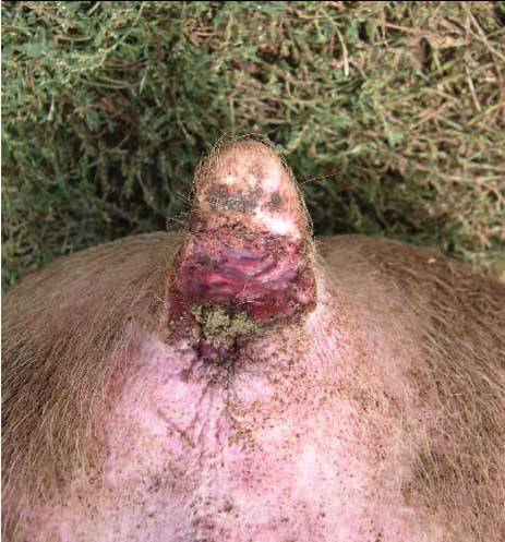
Gross lesions in early affected cases consisted of deep punched-out cavity ulcers commencing to one side of the anal sphincter region (Figure 1). The ulcers had clean, well-circumscribed margins, with rolled edges and minimal obvious granulation. The ulcer floor consisted of a thin necrotic surface and deeper inflammatory reaction. No obvious fistula, abscess, foreign body, tail-biting lesion, prolapse, or other trigger factors were evident in any of the dozens of affected pigs closely examined grossly. The affected pigs (16 to 18 weeks old) were consistently at pubertal onset with fully developed testicles and internal secondary sex glands (eg, seminal vesicles within the lower abdomen). The rectal and colonic feces were regularly of a firm to constipated consistency, with dry matter of 20% to 25%. In over 50% of untreated cases, this initial punched-out ulcer progressed to affect wider and deeper tissues, with evident purulent ulceration and associated inflammation extending across the perineum.
| Figure 1: Punched-out cavity ulcer in a 16-week-old
intact male pig, commencing to one side of the anal sphincter region.
|
All biopsies and autopsy tissue samples at the early stage of ulcerative lesions revealed that deep ulcer tissue consisted of proliferative fibroplasia in a vascular connective tissue collagenous stroma, with scattered neutrophils and macrophages. Histologic Gram stains of tissue samples revealed numerous tissue-located bacteria, predominantly gram-positive coccal forms scattered within the inflammatory stroma. Bacteriologic culture of early cases revealed consistent growth of Streptococcus species, identified as S dysgalactiae by routine API 20 Strep identification system (bio Merieux, Hazelwood, Missouri) in Lancefield Group C, identified by latex bead agglutination reactions using a Streptex kit (Remel Inc, Lenexa, Kansas). This bacteriologic identification was consistent across samples from several groups. Anaerobic cultures also revealed growth of Clostridium perfringens in several cases.
The most consistent features of the outbreaks were the age, origin, and sex of the pigs over numerous pens and barns over a 3-year period. Cases primarily occurred in pigs housed on organic bedding (rice hull and wheat straw combinations), but were occasionally noted in pigs housed on concrete slatted floors. In all forms of housing, however, the onset of outbreaks occurred when pigs were housed in areas of poor water and excreta drainage, with standing fecal-soiled water on the organic or concrete flooring. Concurrently, the pubertal male pigs within affected pig groups were also aggressive and sexually active among their cohorts, and hyperactive when approached.
Discussion
These novel lesions of anal ulceration were clearly
distinguishable from the more common conditions of anal prolapse or
tail-biting lesions by ourselves and attendant farm staff. The tail
itself appeared normal and the anal mucosa showed no noticeable
protrusion throughout individual cases. We suggest that the
proximate cause of the cases was initial trauma to the anus due to
pubertal homosexual behavior with consequent mixed infection,
including
S dysgalactiae and C perfringens, in a water-softened
ano-cutaneous environment. Streptococcus dysgalactiae is
probably a relatively common opportunistic pathogen within
cutaneous ulceration lesions.3 This organism and closely
related organisms are known commensals of the external sexual
organs of pigs.4 Thus, S dysgalactiae may
have been directly introduced into the initial lesion sites from
infected penetrative male genitalia. Whether the particular
isolates of S dysgalactiae in these unusual cases had
altered virulence factors compared to commensal isolates was not
clear from our study.
A number of factors other than the proximate causes may have been involved in these outbreaks. It is possible that the bedding, feces, and urine of these growing pigs may have had the potential to scald the ano-perineum, perhaps due to alterations in pH or cereal content within the diet, allied to poor environmental control of water within pens and barns. However, we also considered an important factor to be the single-source genetic supply of entire and pubertal male pigs of Large White heritage, which showed agitated behavior patterns. Swine veterinary consultants are alerted to this novel and highly deleterious problem in non-castrated male pigs housed on wet organic or other bedding.
References
1. Wilcox CM, Schwartz DA. Idiopathic anorectal ulceration in patients with human immunodeficiency virus infection Am J Gastroenterol. 1994;89:599–604.
2. Toquero L, Briggs CD, Bassuini MM, Rochester JR. Anal ulceration associated with Nicorandil: case series and review of the literature. Colorectal Dis. 2000;8:712–720.
3. Davies CE, Hill KE, Wilson MJ, Stephens P, Hill CM, Harding KG, Thomas DW. Use of 16S ribosomal DNA PCR and denaturing gradient gel electrophoresis for analysis of the microfloras of healing and nonhealing chronic venous leg ulcers. J Clin Microbiol. 2004;42:3549–3557.
4. Bara MR, McGowan MR, O’Boyle D, Cameron RD. A study of the microbial flora of the anterior vagina of normal sows during the different stages of the reproductive cycle. Aust Vet J. 1993;70:256–259.