Diagnostic notes |
Gregory W. Stevenson, DVM, PhD, Diplomate ACVP
Stevenson GW. Brachyspira (Serpulina) pilosicoli and intestinal spirochetosis: How much do we know? Swine Health Prod. 1999;7(6):287-291. Also available in PDF format(248k).
Animal Disease Diagnostic Laboratory and Department of Veterinary Pathobiology, Purdue University, West Lafayette, Indiana 47907-1175; email: greg@addl.purdue.edu.
This diagnostic note has been refereed.  for similar articles.
There are at least five distinct species of Brachyspira (Serpulina) known to infect the large intestine of swine.1-3 Two species are pathogenic:
- Brachyspira hyodysenteriae (formerly Serpulina or Treponema hyodysenteriae), which causes swine dysentery; and
- Brachyspira pilosicoli (formerly Serpulina pilosicoli or Anguillina coli), which causes intestinal spirochetosis.
Three additional species are nonpathogenic:
- Brachyspira innocens, formerly Serpulina or Treponema innocens
- Serpulina intermedia
- Serpulina murdochii
Selected characteristics of each species are summarized in
Table 1.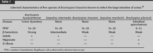
At a light microscopic level, all five of these organisms are morphologically indistinguishable. For this reason, enteric spirochetal diseases in swine can rarely be confirmed by histopathologic examination alone. Specific identification of the pathogen and characteristic lesions are required to confirm a disease diagnosis (see "Diagnosis" section, below).
Why all of the name changes?
In the past 3 decades, there have been significant advances in molecular genetic techniques and a concurrent increase in the number of described spirochetal organisms in swine as well as other mammalian and avian species. This has led to a series of name changes as the phylogenetic relationships of various spirochetes have been established. Until recently, the five characterized species of "serpentine" enteric spirochetes in swine were in the genus Serpulina, including hyodysenteriae, innocens,4,5 pilosicoli,2 intermedia, and murdochii.3 Recently the phylogenetic relationship was clarified between Brachyspira aalborgi, an enteric spirochete found only in humans, and Serpulina hyodysenteriae, innocens, and pilosicoli.6 This was important because both Brachyspira aalborgi and Serpulina pilosicoli infect humans.7-10 Studies demonstrated that all four organisms belonged in the same genus.6 Brachyspira was selected as the genus name based on historic precedent. The genus Brachyspira was established when Brachyspira aalborgi was first described,7 which occurred prior to the establishment of the genus Serpulina.5 Unfortunately, Serpulina intermedia and murdochii were not included in the comparative study. For now, they remain in the genus Serpulina.
Brachyspira pilosicoli
Brachyspira pilosicoli can be presumptively differentiated from other Brachyspira (Serpulina) spp. by culture (weak [beta]-hemolysis) and biochemical testing. Brachyspira pilosicoli is indole negative and hippurate-hydrolysis positive, and lack [beta]-glucosidase activity in the API-ZYM profile.11,12 Definitive identification of B. pilosicoli requires PCR testing.12,13-15 The medium that is most commonly used to culture B. hyodysenteriae in diagnostic laboratories, BJ medium,16 is slightly inhibitory when used to isolate B. pilosicoli, due to the moderate sensitivity of B. pilosicoli to two of the included antibiotics, rifampicin, and spiramycin.17 Culture of B. pilosicoli is most sensitive with a modified BJ media that does not contain rifampicin or spiramycin.
In addition to swine, B. pilosicoli also infects humans,8-10 nonhuman primates,18 dogs,19,20 and several species of birds.21-24 Strains of B. pilosicoli can colonize laboratory mice with fecal shedding for up to 30 days,25 suggesting the potential for rodents to act as reservoirs of infection for swine. Likewise, birds, dogs, and humans are also potential reservoirs for swine. The pathogenic potential of swine strains of B. pilosicoli for humans is unknown, but zoonotic potential exists. Isolation of B. pilosicoli from humans has been associated with clinical disease.8-10 Self-inoculation of a human subject with an avian isolate of B. pilosicoli resulted in mild nausea, abdominal discomfort, and severe headaches.26
Intestinal spirochetosis
Clinical disease
Intestinal spirochetosis is a nonfatal large intestinal disease caused by B. pilosicoli that has been described in field studies of affected swine herds27-31 and in inoculation studies in which disease was reproduced.27,31-33
Clinical disease occurs in weaned pigs primarily 8-16 weeks of age, usually commencing 7-14 days after moving and commingling. This is consistent with the reported incubation period in inoculation studies of 3-16 days,27,31-33 and suggests spread from carrier pigs to susceptible pigs. Typically, the prevalence of disease is 5%-30%, and affected individuals exhibit diarrhea and poor growth for 2-6 weeks. Clinical signs are typically present in a group of pigs for 3-6 weeks. Affected individuals may require up to 28 additional days to reach a slaughter weight of 100 kg (210 lb).34 Uncomplicated intestinal spirochetosis is usually not associated with mortality.35Economic loss is primarily due to reduced growth rate and associated impact on pig flows and market uniformity.31,35 The diarrheic feces are usually first soft and wet with a consistency like "wet cement." Later, feces may change to a watery consistency with a small amount of mucus (i.e., have an "oily" sheen). During recovery or in chronic cases, feces may contain thick tags of mucus. Rarely, flecks of blood may also be present. Affected pigs generally remain alert and active, but appetite is depressed and pigs may show abdominal discomfort and/or may appear gaunt and develop rough hair coats.
Lesions
Gross lesions of intestinal spirochetosis are usually subtle. Pigs are variably gaunt and have rough hair coats. The spiral colon is flaccid, enlarged, and contains abundant watery content with variable amounts of mucus and occasionally some blood. The colonic mesentery and serosa may be thickened by edema in acute cases and the serosa may be thickened by fibrin or fibrous connective tissue in chronic cases (serositis). Colonic lymph nodes are sometimes enlarged. Mucosal lesions are most common and severe in the mid-spiral regions of the spiral, with lesions in the proximal spiral colon the next most prevalent.30 The cecal mucosa is either not involved or has mild lesions.
The colonic and cecal mucosa in affected areas may be congested (reddened) and thickened by edema fluid, forming prominent ridges. Mucosal erosions can occur in variable numbers. With few erosions, the mucosa appears relatively normal (glistening) with a few scattered adherent feed particles. With many erosions, the mucosa appears granular. Fibrin exudation admixed with necrotic cellular debris may result in multifocal fibrinonecrotic tags or plaques. The colonic contents and mucosal surface may contain variable amounts of mucus and occasionally blood. Mucosal lesions are mild compared to classic lesions of swine dysentery or salmonellosis. In order to observe small erosions as evidenced by adherent feed particles or small areas of fibrinonecrotic debris, the mucosa should be gently rinsed free of contents with flowing water. Avoid scraping contents from the mucosa with a postmortem knife, because this will often destroy many of the grossly visible mucosal lesions and alter some microscopic lesions.
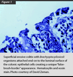 Microscopically, there is a mild to moderately severe
multifocal to diffuse superficial erosive colitis. A variable
amount of fibrinonecrotic debris is on the luminal surface in
areas of erosion. The mucosa is variably thickened by an increased
depth of crypts (crypt hyperplasia), edema of the lamina propria,
and increased numbers of lymphocytes and plasma cells in the lamina
propria and, to a lesser degree, the submucosa. Goblet cell hyperplasia
is common and may cause distention of crypts with mucus. A lesion
unique to B. pilosicoli is end-on attachment of the bacterial
cells to the apical margin of mature epithelial cells on the colonic
luminal surface, creating a "false brush border" or
"hairy" appearance (Figure 1).27,28 Unfortunately,
this lesion is present only inconsistently and only in the early
stages of infection, and cannot be used as a reliable diagnostic
tool. Large serpentine spirochetes typical of Brachyspira
(Serpulina) spp. are more commonly present admixed
with other bacteria in adherent fibrinonecrotic debris in the
superficial lamina propria and in the crypts
Microscopically, there is a mild to moderately severe
multifocal to diffuse superficial erosive colitis. A variable
amount of fibrinonecrotic debris is on the luminal surface in
areas of erosion. The mucosa is variably thickened by an increased
depth of crypts (crypt hyperplasia), edema of the lamina propria,
and increased numbers of lymphocytes and plasma cells in the lamina
propria and, to a lesser degree, the submucosa. Goblet cell hyperplasia
is common and may cause distention of crypts with mucus. A lesion
unique to B. pilosicoli is end-on attachment of the bacterial
cells to the apical margin of mature epithelial cells on the colonic
luminal surface, creating a "false brush border" or
"hairy" appearance (Figure 1).27,28 Unfortunately,
this lesion is present only inconsistently and only in the early
stages of infection, and cannot be used as a reliable diagnostic
tool. Large serpentine spirochetes typical of Brachyspira
(Serpulina) spp. are more commonly present admixed
with other bacteria in adherent fibrinonecrotic debris in the
superficial lamina propria and in the crypts 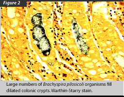 (Figure 2).
Unfortunately, B. pilosicoli cannot be differentiated from
other Brachyspira (Serpulina) spp. based only on light
microscopic morphology. Apart from the unique but inconsistent
lesion of end-on attachment by B. pilosicoli, the microscopic
lesions of intestinal spirochetosis are relatively nonspecific
and can be mimicked by mild lesions of salmonellosis or swine
dysentery.
(Figure 2).
Unfortunately, B. pilosicoli cannot be differentiated from
other Brachyspira (Serpulina) spp. based only on light
microscopic morphology. Apart from the unique but inconsistent
lesion of end-on attachment by B. pilosicoli, the microscopic
lesions of intestinal spirochetosis are relatively nonspecific
and can be mimicked by mild lesions of salmonellosis or swine
dysentery.
Epidemiology
Knowledge of the epidemiology of intestinal spirochetosis is limited, based on few inoculation studies27,31-33 and field studies.26-30,34,36,37 Infection with B. pilosicoli has been reported in swine in nearly every country with a significant swine industry. The proportion of infected swine herds in the United States is unknown. In a limited study of diarrheic pigs on 10 grower sites in a single United States swine production company, B. pilosicoli was isolated in 50% of sites (Duhamel GE, et al. Proc Am Assoc Vet Lab Diagn. 1996; 45). In a study of 85 swine herds with a history of colitis in the United Kingdom, B. pilosicoli was detected in 52% and was the sole pathogen detected in 33%.30 In Sweden, a study of 894 farms observed B. pilosicoli in 18% of farms.37 In Finland, in a study of 50 finishing sites stocked from farrowing sites of "LSO 2000 quality chain" health status, investigators detected B. pilosicoli in 28% of farms (Heinonen M. Proc IPVS Cong. 1998;2:57).
Transmission of B. pilosicoli is thought to be exclusively fecal-oral. The greatest risk factor for infection of negative pigs is exposure to fresh feces from shedding carrier pigs. Brachyspira pilosicoli survives in lake water for 66 days at 4 degrees C.26 It is likely that B. pilosicoli, like B. hyodysenteriae, survives in anaerobic lagoons and in moist fecal matter. Therefore, flush gutters using recycled lagoon water, inadequate cleaning of feces from pens/facilities, and fecal contamination in trucks/trailers. should all be considered significant risk factors for infection. Other species known to sometimes carry B. pilosicoli--including humans, dogs, birds, and possibly mice--may pose some biosecurity risk to negative herds. Bird-proofing buildings and controlling rodents are recommended as prudent preventive measures. Fecal contamination by feral birds of water sources used for drinking or flushing gutters is also a potential source of B. pilosicoli.26
Not all pigs that are infected with B. pilosicoli develop diarrhea. In oral inoculation studies, nearly all pigs become colonized and shed B. pilosicoli in feces, but only 30%-70% develop diarrhea.27,31-33 Subclinically infected pigs may develop typical gross and microscopic lesions of intestinal spirochetosis.31 Inoculated pigs remain colonized and shed B. pilosicoli in feces for up to 6 weeks.31 Exposure dose and risk of disease presumably increase with management practices that increase exposure to carrier animals or contaminated feces, including:
- frequent mixing of pigs,
- continuous pig flows,
- poor ventilation/sanitation, and
- high stocking densities.
Pelleted feed appears to increase the risk of diarrhea in B. pilosicoli-infected pigs (MacDougald D. Proc AASP Ann Meet. 1997;497).36 When the ration is changed from pellets to meal, the proportion of diarrheic pigs typically decreases.
Diagnosis
A definitive diagnosis of intestinal spirochetosis requires observation of typical colonic lesions and confirmation of infection by B. pilosicoli. Other diseases that should be excluded by testing are salmonellosis, swine dysentery, proliferative enteritis, whipworm infestation, and possibly yersiniosis caused by Yersinia pseudotuberculosis.30 Tests available to detect infection with B. pilosicoli include culture and/or PCR. Presumptive identification of B. pilosicoli is possible based on weak-[beta]-hemolysis of colonies on blood-agar and results of biochemical testing.11,12 Definitive identification of B. pilosicoli requires PCR testing.11,13-15 Tests to detect B. pilosicoli are most sensitive when conducted on colonic and cecal mucosal samples collected from euthanized pigs. Samples should be immediately chilled and shipped on ice overnight to the laboratory for testing. Fecal samples may be used to confirm infection and predict prevalence of infection in populations. However, sensitivity for either culture or PCR testing is lower in fecal samples as compared to colonic or cecal mucosal samples from the same pigs. Duhamel estimated that the sensitivity of fecal culture for B. pilosicoli is approximately 80% under ideal conditions when pigs are shedding large numbers of organisms with no antibiotics in the feed (Duhamel GE, et al. Proc IPVS Cong. 1998; 3:141). Under most field conditions, the sensitivity of fecal culture is significantly lower.
Fecal samples for culture should be collected on swabs and immersed in Amies transport medium with activated charcoal (Bioport 4(TM), Precision Dynamics Corp; San Fernando, California), chilled, and shipped on ice overnight to the laboratory for testing. In-situ hybridization testing for B. pilosicoli done on formalin-fixed sections of colon has been described experimentally as a sensitive and specific method for diagnosing intestinal spirochetosis, but is not yet available in diagnostic laboratories in the United States (Jensen TK. Proc IPVS Cong. 1998;2:58).
To adequately determine the cause of diarrhea in a population of grower pigs:
- acutely affected pigs should be euthanized and examined by necropsy. The entire gastrointestinal tract should be examined. Special care should be taken to open and examine the entire ileum, cecum, and colon. The mucosa should be gently rinsed with water (not scraped with a knife) during examination and prior to collecting samples. Minimally, fresh-chilled and formalin-fixed samples of jejunum, ileum, proximal and mid-spiral colon, cecum, and mesenteric/colonic lymph nodes should be submitted for bacteriology, histopathology, and virology (jejunum and ileum for TGEV and rotaviruses).
- Individual or pooled fecal samples from acutely affected animals should also be submitted chilled (not frozen) for EM to detect viruses including transmissible gastroenteritis virus and rotaviruses.
- Fecal samples from a statistically representative sample of the population (20-30) should be collected in Amies transport medium with activated charcoal, chilled, and transported on ice for PCR testing and/or culture for Brachyspira (Serpulina) spp., Lawsonia intracellularis, and Salmonella spp.
Treatment and prevention
Brachyspira pilosicoli is generally sensitive to the same antibiotics as B. hyodysenteriae. Variable clinical response of intestinal spirochetosis to treatment with antibiotics is described (MacDougald D. Proc AASP Ann Meet. 1997;497).29,33,34,38 In a study of 19 United States strains of B. pilosicoli, all were susceptible by in vitro testing to carbadox and tiamulin, 47% were susceptible to gentamycin, and 42% were susceptible to lincomycin.38 Most schemes for control of intestinal spirochetosis combine therapeutic concentrations of antimicrobials during the first few weeks that pigs are in grower buildings, in addition to sanitation measures; i.e., cleaning and disinfecting pens/buildings between groups of pigs. It is assumed, but not proven, that schemes combining treatment and sanitation for the elimination of B. hyodysenteriae would also be effective against B. pilosicoli. Like B. hyodysenteriae, B. pilosicoli appears to become resistant to antibiotics over time and producers should rotate the use of antibiotics.
Studies to determine whether it would be cost effective to eliminate B. pilosicoli have not been undertaken. Vaccinating pigs with an experimental formalin-killed whole cell bacterin with Freund's incomplete adjuvant did not protect pigs from infection or disease after experimental challenge with B. pilosicoli (Hampson DJ, et al. Proc IPVS Cong. 1998;2:56). In the same study, nonvaccinated control pigs were colonized, and developed lesions and clinical disease, yet did not mount a significant humoral immune response (La T, et al. Proc IPVS Cong. 1998;3:131).
What we don't yet know
As is true of many diseases, especially recently recognized or emerging diseases, some of the most practical and important questions remain unanswered:
- What is the prevalence of B. pilosicoli infection in the United States swine herd?
- In an infected herd, what proportion of pigs become infected and when?
- What is the cost of subclinical infection?
- What is the cost of clinical disease?
- What are the best methods of treatment, control, or elimination? Are they cost effective?
- Is infection with other enteric agents additive or synergistic?
- In diarrheal disease in which multiple pathogenic agents are demonstrated in a population, what is the relative contribution of each?
- What will the name of Brachyspira pilosicoli be changed to next?
References
1. Lee JI, Hampson DJ, Lymbery AJ, Harders SJ. The porcine intestinal spirochaetes: Identification of new genetic groups. Vet Microbiol. 1993;34:273-285.
2. Trott DJ, Stanton TB, Jensen NS, Duhamel GE, Johnson JL, Hampson DJ. Serpulina pilosicoli sp. nov., the agent of porcine intestinal spirochetosis. Int J Sys Bacteriol. 1996; 46:206-215.
3. Stanton TB, Fournie-Amazouz E, Postic D, Trott DJ, Grimont PAD, Barantan G, Hampson DJ, Saint Girons I. Recognition of two new species of intestinal spirochetes: Serpulina intermedia sp. nov. and Serpulina murdochii sp. nov. Int J Sys Bacteriol. 1997;47:1007-1012.
4. Stanton TB, Jensen NS, Casey TA, Tordoff LA, Deewhirst FE, Paster BJ. Reclassification of Treponema hyodysenteriae and Treponema innocens in a new genus, Serpula gen. nov., as Serpula hyodysenteriae comb. nov. and Serpula innocens comb. nov. Int J Syst Bacteriol. 1991;41:50-58.
5. Stanton TB. Proposal to change the genus designation Serpula to Serpulina gen. nov. containing the species Serpulina hyodysenteriae comb. nov. and Serpulina innocens comb. nov. Int J Syst Bacteriol. 1992;42:189-190.
6. Ochiai S, Adachi Y, Mori K. Unification of the genera Serpulina and Brachyspira, and proposals of Brachyspira hyodysenteriae comb. nov., Brachyspira innocens comb. nov. and Brachyspira pilosicoli comb. nov. Microbiol Immunol. 1997;41:445-452.
7. Hovind-Hougen K, Birch-Andersen A, Henrik-Nielsen R, Orholm M, Pedersen JO, Teglbajaerg PS, Thaysen EH. Intestinal spirochetosis: Morphological characterization and cultivation of the spirochete Brachyspira aalborgi gen. nov., sp. nov. J Clin Microbiol. 1982;16:1127-1136.
8. Trott DJ, Jensen NS, Saint Girons I, Oxberry SL, Stanton TB, Lindquist D, Hampson DJ. Identification and characterization of Serpulina pilosicoli isolates recovered from the blood of critically ill patients. J Clin Microbiol. 1997;35:482-485.
9. Trott DJ, Combs BG, Mikosza ASJ, Oxberry SL, Robertson ID, Passey M, Taime J, Sehuko R, Alpers MP, Hampson DJ. The prevalence of Serpulina pilosicoli in humans and domestic animals in the Eastern Highlands of Papua New Guinea. Epidemiol Infect. 1997;119:369-379.
10. Trivett-Moore NL, Gilbert GL, Law CLH, Trott DJ, Hampson DJ. Isolation of Serpulina pilosicoli from rectal biopsy specimens showing evidence of intestinal spirochetosis. J Clin Microbiol. 1998;36:261-265.
11. Fellstrom C, Pettersson B, Thomson J, Gunnarsson A, Persson M, Johansson KE. Identification of Serpulina species associated with porcine colitis by biochemical analysis and PCR. J Clin Microbiol. 1997;35:462-467.
12. Fellstrom C, Gunnarsson A. Phenotypical characterisation of intestinal spirochaetes isolated from pigs. Res Vet Sci. 1995;58:1-4.
13. Park NY, Chung CY, McLaren AJ, Atyeo RF, Hampson DJ. Polymerase chain reaction for identification of human and porcine spirochaetes recovered from cases of intestinal spirochaetosis. FEMS Microbiol Letters. 1995; 125:225-230.
14. Muniappa N, Mathiesen MR, Duhamel GE. Laboratory identification and enteropathogenicity testing of Serpulina pilosicoli associated with porcine colonic spirochetosis. J Vet Diagn Invest. 1997;9:165-171.
15. Leser TD, Moller K, Jensen TK, Jorsal SE. Specific detection of Serpulina hyodysenteriae and potentially pathogenic weakly [beta]-haemolytic porcine intestinal spirochetes by polymerase chain reaction targeting 23S rDNA. Mol Cell Probes. 1997;11:363-372.
16. Kunkle RA, Kinyon JM. Improved selective medium for the isolation of Treponema hyodysenteriae. J Clin Microbiol. 1988;26:2357-2360.
17. Trott DJ, Stanton TB, Jensen NS, Hampson DJ. Phenotypic characteristics of Serpulina pilosicoli, the agent of porcine intestinal spirochetosis. FEMS Microbiol Lett. 1996; 142:209-214.
18. Duhamel GE, Elder RO, Muniappa N, Mathiesen MR, Wong VJ, Tarara RP. Colonic spirochetal infection in nonhuman primates that were associated with Brachyspira aalborgi, Serpulina pilosicoli, and unclassified flagellated bacteria. Clin Infect Dis. 1997;25(Suppl 2):S186-S188.
19. Duhamel GE, Muniappa N, Mathiesen MR, Johnson JL, Toth J, Elder RO, Doster AR. Certain canine weakly beta-hemolytic intestinal spirochetes are phenotypically and genotypically related to spirochetes associated with human and porcine intestinal spirochetosis. J Clin Microbiol. 1995;33:2211-2215.
20. Duhamel GE, Trott DJ, Muniappa N, Mathiesen MR, Tarasiuk K, Lee JI, Hampson DJ. Canine intestinal spirochetes consist of Serpulina pilosicoli and a newly identified group provisionally designated "Serpulina canis" sp. nov. J Clin Microbiol. 1998;36:2264-2270.
21. Trampel DW, Jensen NS, Hoffman LJ. Cecal spirochetosis in commercial laying hens. Avian Diseases. 1994;38:895-898.
22. McLaren AJ, Trott DJ, Swayne DE, Oxberry SL, Hampson DJ. Genetic and phenotypic characterization of intestinal spirochetes colonizing chickens and allocation of known pathogenic isolates to three distinct genetic groups. J Clin Microbiol. 1997;35:412-417.
23. Swayne DE, McLaren AJ. Avian intestinal spirochaetes and avian intestinal spirochaetosis. In: Hampson DJ, Stanton TB, eds. Intestinal spirochaetosis in domestic animals and humans. Wallingford, UK: CAB International. 1997;267-300.
24. Webb DM, Duhamel GE, Mathiesen MR, Muniappa N, White AK. Cecal spirochetosis associated with Serpulina pilosicoli in captive juvenile ring-necked pheasants. Avian Dis. 1997;41:997-1002.
25. Sacco RE, Trampel DW, Wannemuehler MJ. Experimental infection of C3H mice with avian, porcine, or human isolates of Serpulina pilosicoli. Infect Immun. 1997; 65:5349-5353.
26. Oxberry SI, Trott DJ, Hampson DJ. Serpulina pilosicoli, waterbirds and water: Potential sources of infection for humans and other animals. Epidemiol Infect. 1998;121:219-225.
27. Taylor DJ, Simmons JR, Laird HM. Production of diarrhoea and dysentery in pigs by feeding pure cultures of a spirochaete differing from Treponema hyodysenteriae. Vet Rec. 1980;106:326-332.
28. Jacques M, Girard C, Higgins R, Goyette G. Extensive colonization of the porcine colonic epithelium by a spirochete similar to Treponema innocens. J Clin Micro. 1989; 27:1139-1141.
29. Girard C, Lemarchand T, Higgins R. Porcine colonic spirochetosis: A retrospective study of eleven cases. Can Vet J. 1995;36:291-294.
30. Thomson JR, Smith WJ, Murray BP. Investigations into field cases of porcine colitis with particular reference to infection with Serpulina pilosicoli. Vet Rec. 1998;142:235-239.
31. Duhamel GE. Colonic spirochetosis caused by Serpulina pilosicoli. Large An Pract. 1998;19:14-22.
32. Thomson JR, Smith WJ, Murray BP, McOrist S. Pathogenicity of three strains of Serpulina pilosicoli in pigs with a naturally acquired intestinal flora. Infect Immun. 1997;65:3693-3700.
33. Trott DJ, Huxtable CR, Hampson DJ. Experimental infection of newly weaned pigs with human and porcine strains of Serpulina pilosicoli. Infec Immun. 1996;64:4648-4654.
34. Taylor DJ , Trott DJ. Porcine intestinal spirochaetosis and spirochaetal colitis. In: Hampson DJ, Stanton TB, eds. Intestinal Spirochaetosis in Domestic Animals and Humans. Wallingford, UK: CAB International; 1997;211-241.
35. Hampson DJ, Trott DJ. Spirochetal diarrhea / Porcine intestinal spirochetosis. In: Staw B, D'Allaire S, Mengeling W, Taylor D, eds. Diseases of Swine. 8th edition. Ames, Iowa: Iowa State University Press; 1999:553-562.
36. Spearman JG, Nayar G, Sheridan M. Colitis associated with Treponema innocens in pigs. Can Vet J. 1988; 29:747.
37. Fellstrom C, Pettersson B, Johansson KE, Lundeheim N, Gunnarsson A. Prevalence of Serpulina species in relation to diarrhea and feed medication in pig rearing herds in Sweden. Am J Vet Res. 1996;57:807-811.
38. Duhamel GE, Kinyon JM, Mathiesen MR, Murphy DP, Walter D. In vitro activity of four antimicrobial agents against North American isolates of porcine Serpulina pilosicoli. J Vet Diagn Invest. 1998;10:448-354.