| Original research | Brief Communication |
Cite as: Dominguez BJ, Duckworth LA, Jones ML. Comparison of regional limb injection to systemic medication for the treatment of septic lameness in female breeding swine. J Swine Health Prod. 2016;24(2):93–96.
Also available as a PDF.
SummaryTwenty female breeding swine with acute septic lameness received lincomycin systemically or via regional limb perfusion (RLP). There was no significant difference in the time to healing between methods. However, lameness resolved earlier in a numerically higher proportion of subjects receiving RLP than systemic treatment. | ResumenVeinte cerdas con cojera séptica aguda recibieron lincomicina de forma sistémica o vía perfusión de miembro regional (RLP por sus siglas en inglés). No hubo una diferencia significativa en el tiempo de curación entre los dos métodos. Sin embargo, la cojera se resolvió más rápido en una proporción numérica más alta en los sujetos que recibieron RLP que con el tratamiento sistémico.
| ResuméVingt truies souffrant de boiterie septique aigüe ont reçu de la lincomycine par voie systémique ou via une perfusion régionale du membre (PRM). Il n’y avait pas de différence significative dans le temps de guérison entre les deux méthodes. Toutefois, la boiterie s’est résolue plus rapidement dans une proportion plus élevée de sujets recevant le traitement PRM que le traitement systémique. |
Keywords: swine, regional limb perfusion, septic arthritis
Search the AASV web site
for pages with similar keywords.
Received: February 10, 2015
Accepted: September 29, 2015
Lameness in swine is a topic of animal welfare and economic concern.1 Lameness is an important cause of involuntary culling, with rates as high as 15%,2 and infectious arthritis has been reported to be the second most important cause of lameness in culled sows.3 Regional limb perfusion (RLP) with an antimicrobial is used in bovine and equine species for the treatment of distal limb infections. Using a tourniquet to isolate a region of the limb, RLP delivers the antimicrobial from the vasculature to the surrounding tissue via diffusion.4,5 As an alternative to systemic antimicrobial therapy, regional limb perfusion results in higher local drug concentrations for an extended period of time and decreases drug dose, systemic concentrations, adverse drug effects (potentially), number of treatments, convalescent time, and labor.6 Conventional treatment of septic lameness in swine consists of systemic administration of antimicrobials, with lincomycin the only antimicrobial labelled for the treatment of lameness in swine.
The purpose of this study was to introduce and evaluate the efficacy of RLP of lincomycin as an alternative treatment for septic lameness in swine.
Materials and methods
This study was reviewed and approved by the Texas A&M University Institutional Animal Care and Use Committee.
Patient selection and observation
Two sow farms owned by a single client were utilized as sources of animals for this study. Cases were selected as gilts and sows were being loaded into farrowing crates or as they moved about gestation pens. Gestation pens measured 6.0 m × 4.5 m and housed 6 to 10 sows, and farrowing crates measured 0.7 m × 2.4 m. All pigs in each cohort of breeding groups were issued a lameness grade based on the Zurbrigg and Blackwell scale,7 with Grade 1 categorized as not lame; 2, lame; and 3, unable to ambulate. Subjects that scored a Grade 2 with acute septic lameness localized to the distal limb or foot were included in this study. Acute septic lameness was defined by the presence of swelling and heat in the metatarsophalangeal or interphalangeal joints and associated soft tissue structures. Animals with chronic lesions, characterized by the presence of exuberant granulation tissue or bony prominences, were not included in this study. Animals were enrolled in the study over a 10-week period.
Treatment
Twenty animals were identified as lame. Selected subjects were randomly assigned to treatment groups using a random number generator. Nine animals were treated systemically as controls, and 11 animals were treated via RLP. No animal identified as lame due to septic arthritis remained untreated.
Control group treatment. The control group received once-daily systemic treatments of Lincomycin HCL (300 mg per mL; Pharmacia and Upjohn Co, Kalamazoo, Michigan) at a dose of 11 mg per kg intramuscularly (IM) in the neck on 3 consecutive days.
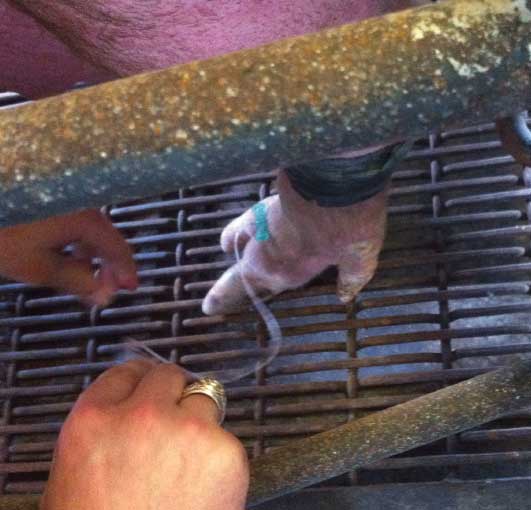
Regional intravenous limb perfusion. Regional intravenous limb perfusion was performed by restraining the animal using a snare, then applying a 3.75-cm wide rubber tourniquet to the mid-metacarpal or metatarsal region of the affected limb. The tourniquet was fabricated by splitting a 26-inch bicycle inner tube (Bell Sports, Rantoul, Illinois) in half. This allowed sufficient length for the tourniquet to be wrapped around the leg approximately three times and secured under itself. Animals were completely washed prior to examination, and the area between the toes of the affected foot was wiped three times with alcohol, allowing time for drying. A 21-gauge butterfly catheter (Terumo Corporation, Tokyo, Japan) was inserted into the dorsal common digital vein at a point approximately 1.3 cm proximal to the interdigital cleft (Figure 1). For RLP, 100 mg of Lincomycin HCL (0.3 mL) was diluted to 3 mL with 0.9% sterile saline and administered through the catheter, followed by a flush of air just sufficient to clear the catheter (extra-label use). The catheter was then removed and the animal was released from the snare. The tourniquet was left in place for 30 minutes while the sow was kept in her farrowing crate or in a small holding pen. This procedure was repeated once daily for 3 days to mimic the label dosing of systemic lincomycin.
Figure 1: Placement of needle for regional limb perfusion with lincomycin to treat septic arthritis in a sow. A 21-gauge butterfly catheter (Terumo Corporation, Tokyo, Japan) was inserted into the dorsal common digital vein at a point approximately 1.3 cm proximal to the interdigital cleft.

Treatment evaluation. Animals were observed in their crate or pen once a week for 4 weeks, beginning immediately following treatment. Animals were observed by one or more of the authors, with one author (BJD) being involved in all observations. Any evidence of lameness was noted, particularly in the originally affected limb, including ability to rise and hesitancy to place the affected foot on the ground. Feet were palpated for evidence of heat or swelling. The same Zurbrigg and Blackwell scoring system7 was utilized throughout the post-treatment observations.
Statistical analysis
Descriptive statistics were determined for parity and separated by location and treatment. The proportion of animals responding to treatment each week was determined by location and treatment. Univariable logistic regression for each week post treatment determined the change in odds of resolution of lameness by route of administration, location in barn, body weight, parity, and leg affected.
Results
Placement of the tourniquet was well tolerated in all patients, as evidenced by no vocalization and very brief retraction of the leg before resuming normal posture. There was typically a brief reaction to needle placement exhibited by kicking or lifting the leg. The total time for treatment via regional limb perfusion was approximately 35 minutes, while the hands-on time was less than 5 minutes per animal. Two sows prematurely lost their tourniquets during one treatment each at 9 and 15 minutes post injection. In all but one animal, the dorsal common digital vein was easily accessed. That animal received systemic treatment and was excluded from the study.
The desired treatment outcome measured was complete resolution of lameness by each observational period. There was no significant difference between systemic and RLP routes on any day of evaluation. Results are summarized in Table 1.
Table 1: Resolution of lameness in sows with septic arthritis of the distal limb or foot and treated with lincomycin systemically or via regional limb perfusion (RLP)*
| No. (%) of sows that achieved complete resolution of lameness | ||||
|---|---|---|---|---|
| Day† | 7 | 14 | 21 | 28 |
| Systemic | 4/9 (44.4) | 6/9 (66.7) | 8/9 (88.9) | 8/9 (88.9) |
| RLP | 5/11 (45.5) | 8/11 (72.7) | 9/11 (81.8) | 7/9 (77.8) |
| Total | 9/20 (45.0) | 14/20 (70.0) | 17/20 (85.0) | 15/18 (83.3) |
* A total of 20 sows were selected from two farms in a single production system. Sows were observed while loading into farrowing crates or while in gestation pens. Lameness was graded as 1 (not lame), 2 (lame), or 3 (unable to ambulate).7 Controls (n = 9) were treated with lincomycin systemically (11 mg/kg intramuscularly on 3 consecutive days) and treatment sows (n = 11) were treated with 100 mg lincomycin via RLP, with the tourniquet left in place for 30 minutes, on 3 consecutive days.
† Day post initial treatment. Sows were enrolled in the study over a 10-week period. Two RLP sows were culled for reproductive reasons before the trial ended. Univariable logistic regression for each week post treatment was used to determine the change in odds of resolution of lameness by route of administration, location in barn, body weight, parity, and leg affected. There was no significant difference in lameness scores between systemic and RLP routes on any day of evaluation.
Among animals in the farrowing barn treated with RLP, lameness in 59% improved to Grade 1 by day 7, and 83.3% of animals showed the same improvement from day 14 onwards. Among animals in the farrowing barn administered systemic treatment, 80% improved to Grade 1 by day 7 and 100% by day 14. In gestation, no improvement in lameness grade was noted in the animals administered systemic treatment until day 14, when one of four was Grade 1, and three of the four improved to Grade 1 by day 21. Among the RLP group in gestation, 60% and 80% of animals had improved to Grade 1 by day 7 and day 21, respectively. Two animals treated by RLP in gestation had improved to Grade 1 by day 21, but were culled for reproductive reasons prior to the end of the study.
Univariable logistic regression analysis demonstrated a trend toward placement in a farrowing barn promoting resolution at 7 days (P = .08). At 14 days, housing in a farrowing barn significantly promoted resolution of lameness compared to housing in gestation (P = .04). No other factor (route, leg affected, bodyweight, or parity) was significant in univariable analysis.
Post-hoc power analysis found the power of this study much less than expected (5.02%). Given the observed proportions of treatment success by each route, it is now estimated that 950 animals would be needed in each group to obtain a power of 80%.
Discussion
The difference that was observed between systemic and RLP treatments was not as great as expected. According to farm managers, the efficacy of systemic treatment reported on the farm was lower than experienced during the study. This may be an effect of decreased lincomycin use on the farm leading to increased susceptibility, a misconception concerning the original efficacy of the antimicrobial on the farm, or improved early diagnosis of lameness by researchers. In gestation pens, it took 3 weeks for a majority of sows and gilts to show resolution of the lameness. This time frame might be longer than farm-manager expectations, which may have resulted in premature culling.
Cases were targeted for acute signs of lameness. Enrollment during the transition to farrowing barns was attempted, as animals could be observed ambulating and then were placed into individual farrowing crates which facilitated treatment. Allowing sows to stay individually penned during a time when they are naturally less active, around farrowing, may have additionally enhanced the healing process. All animals identified as Grade 2 lame were treated for their individual welfare. Inclusion of a non-treated control group of Grade-2-lame animals would have helped determine the proportion of animals that resolved without treatment.
This study has revealed that RLP is a feasible method of treating lameness in individual animals. While often placed without visualization of the vein, the butterfly catheter was easily placed in the dorsal common digital vein with the aid of a tourniquet. The results indicate that in some situations, RLP may provide more rapid resolution of septic causes of lameness, and may be a useful alternative for treatment of lameness in individual animals.
Regional intravenous limb perfusion in swine requires some technical skill, comparable to administering any intravenous injection. The area that can be treated in this manner in swine is small compared to that in other large animals because of their anatomy. In one sow, the catheter could not be placed, and premature tourniquet loss occurred in two animals. Tourniquet loss did not appear to have a negative effect on resolution of lameness in these subjects.
The pharmacokinetics of drugs administered via RLP is imperfectly understood, particularly in swine. The dose for this study was selected to represent a reasonable reduction from the systemic dose, similar to the study reported by Navarre et al.6 Gilliam et al8 attempted to quantify the dose more accurately by weighing cattle legs cut at the level of the tourniquet and calculating the dose on the basis of that weight. Both approaches provide only efficient estimates of an appropriate dose, and both methodologies result in doses that are much less than the total systemic dose. For this study, the tourniquet was kept in place for 30 minutes, but the actual time that is needed is not known. The amount of time required to allow the drug to reach adequate tissue concentrations has not been determined in swine. Principles guiding the prudent use of antimicrobials in food-producing animals require justification to allow for extra-label use, including RLP. This study suggests that the resolution of lameness may be more rapid with RLP, resulting in a more rapid improvement in welfare. Because the antimicrobial is being given in an extra-label manner, by regulation, the withdrawal time must be extended by a reasonable amount. Practitioners need to be especially cognizant of prohibited drugs and drugs voluntarily banned for food animals that may be administered by RLP in other species. Practitioners also need to be aware of the laws governing their area of practice, as regulations vary by country.
Implications
• Reducing the use of antimicrobials in food-producing animals may be achieved through more widespread use of RLP.
• Regional intravenous limb perfusion of an antimicrobial to treat lameness is feasible in swine.
Acknowledgements
Supported by a grant from the American Association of Swine Veterinarians Foundation.
Conflict of interest
None reported.
Disclaimer
Scientific manuscripts published in the Journal of Swine Health and Production are peer reviewed. However, information on medications, feed, and management techniques may be specific to the research or commercial situation presented in the manuscript. It is the responsibility of the reader to use information responsibly and in accordance with the rules and regulations governing research or the practice of veterinary medicine in their country or region.
References
1. Smith B. Lameness in pigs associated with foot and limb disorders. In Pract. 1988;10:113–117.
2. USDA National Animal Health Monitoring Service. Swine 2006 Part I: Reference of Swine Health and Management Practices in the United States. 2007. Available at https://www.aphis.usda.gov/animal_health/nahms/swine/downloads/swine2006/Swine2006_dr_PartI.pdf. Accessed 30 October 2015.
3. Dewey CE, Friendship RM, Wilson MR. Clinical and postmortem examination of sows culled for lameness. Can Vet J. 1993;34:555–556.
4. Whitehair KJ, Blevins WE, Fessler JF, Sickle DC, White MR, Bill RP. Regional perfusion of the equine carpus for antibiotic delivery. Vet Surg. 1992;21:279–285.
5. Finsterbusch A, Argaman M, Sacks T. Bone and joint perfusion with antibiotics in the treatment of experimental staphylococcal infection in rabbits. J Bone Joint Surg Am. 1970;52:1424–1432.
6. Navarre CB, Zhang L, Sunkara G, Duran SH, Kompella UB. Ceftiofur distribution in plasma and joint fluid following regional limb injection in cattle. J Vet Pharm Therap. 1999;22:13–19.
7. Zurbrigg K, Blackwell T. Injuries, lameness, and cleanliness of sows in four group-housing gestation facilities in Ontario. J Swine Health Prod. 2006;14:202–206.
8. Gilliam JN, Streeter RN, Papich MG, Washburn KE, Payton ME. Pharmacokinetics of florfenicol in serum and synovial fluid after regional intravenous perfusion in the distal portion of the hind limb of adult cows. Am J Vet Res. 2008;69:997–1004.